
Medical Specialties
Medical Specialties
Explore a comprehensive collection of scientific and clinical resources tailored for HCPs, including peer insights, clinical case studies, and symposia. Designed for neurosurgeons, ophthalmologists, and specialists in Plastic and Reconstructive surgery, ENT, and dentistry. This collection highlights the latest advancements in surgical microscopy. Discover how cutting-edge surgical technologies, such as AR fluorescence, 3D visualization, and intraoperative OCT imaging, empower confident decision-making and precision in complex surgeries.

Find Relevant Specimen Details from Overviews
Switch from searching image by image to seeing the full overview of samples quickly and identifying the important specimen details instantly with confocal microscopy. Use that knowledge to set up…

Benefits of Combining STED and Lifetime
In this interview, Professor Alberto Diaspro talks about the advantages of the White Light Laser and the TauSTED capabilities of STELLARIS 8 STED. He speaks about his experience with the confocal…

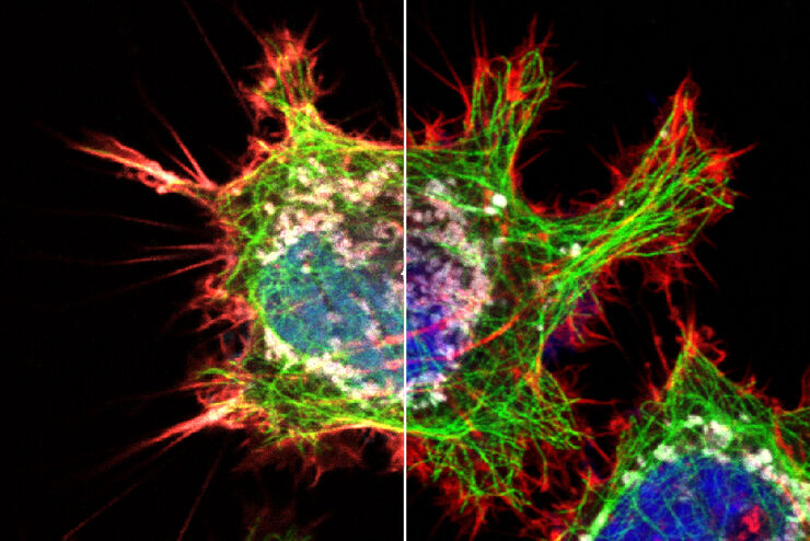
Live Cell Imaging Gallery
Live cell microscopy techniques are fundamental to get a better understanding of cellular and molecular function. Today, widefield microscopy is the most common technique used to visualize cell…

Tissue Image Gallery
Visual analysis of animal and human tissues is critical to understand complex diseases such as cancer or neurodegeneration. From basic immunohistochemistry to intravital imaging, confocal microscopy…
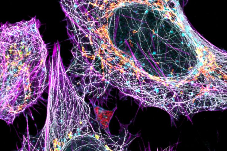
Multicolor Image Gallery
Fluorescence multicolor microscopy, which is one aspect of multiplex imaging, allows for the observation and analysis of multiple elements within the same sample – each tagged with a different…
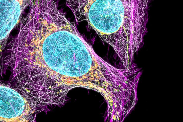
Cell Biology Image Gallery
Cell biology studies the structure, function and behavior of cells, including cell metabolism, cell cycle, and cell signaling. Fluorescence microscopes are an integral part of a cell biologist…
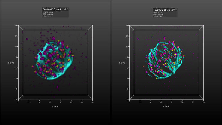
Kinetochore Assembly during Mitosis with TauSTED on 3D
Three-dimensional organization of the mitotic spindle together with the distribution of CENP-C and BUB1 based on TauSTED with multiple STED lines (592, 660 and 775 nm) can provide insights…
Explore Innovative Techniques to Separate Fluorophores with Overlapping Spectra
In this article we explore several strategies you can take to improve the separation of fluorophores and increase the number of fluorescent probes you can distinguish in your sample.

STELLARIS White Light Lasers
When it comes to choosing fluorescent probes for your multi-color experiments, you shouldn’t have to compromise. Now you can advance beyond conventional excitation sources that limit your fluorophore…
