Introduction
Dr. Parolini shares her opinion about the value of intraoperative OCT and the benefits of the technology for daily practice.
Advantages of intraoperative OCT in surgery
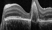
In this video segment Dr. Parolini explains how supplementing the microscope view with OCT imaging results in more information for the surgeon. According to Dr. Parolini the additional sub-surface details allow surgeons to work more precisely and can help to speed up procedures.
Application examples
A selection of cases, where intraoperative OCT was useful during surgery and even influenced the surgery itself.
Membrane peelings
OCT is particularly helpful in complicated cases of membrane peelings where there may be low contrast due to low choroidal coloring, choroidal atrophy, retinal edema, drusen, or microhemorrhages. These factors can make it difficult to see all the necessary detail with the microscope alone.
Dr. Parolini shares some examples of membrane peeling cases in this section of the webinar. She explains how intraoperative OCT supported her by providing a precise view and more visual information, in order to:
- Avoid inducing a macular hole
- Get indication of how much needs to be peeled
- Decide at which point to stop the peeling
Retinal detachment
Dr. Parolini shares her experience in retinal detachment cases, emphasizing how intraoperative OCT supported her surgical assessment, for example when:
- Judging if the retina is reattached, and if there’s subretinal fluid
- Deciding whether to peel the internal limiting membrane (IML) or not
- Differentiating areas of detachment and areas of schisis.
The cases include:
- PFCL and air injection to attach the retina, checking how much fluid remains under the retina
- IML peeling in a case with very thin fovea, where Dr. Parolini changed her plans intraoperatively in order to avoid inducement of a full thickness macular hole (FTMH)
- A specific type of a retinal detachment in a congenital retinoschisis where it was difficult to differentiate areas of real detachments vs. areas of retina schisis
Macular buckle
In this part of the webinar Dr. Parolini discusses the types of Myopic Traction Maculopathy where a macular buckle is part of the treatment. She describes how she used intraoperative OCT to confirm the exact location of the buckle and the indentation level of the sclera. This section also provides a detailed overview table from Dr. Parolini categorizing all types of myopic traction maculopathies.
Transplant of choroid
Choroidal transplants are a treatment for patients with different types of macular apathies that do not respond to other treatment.
In the following video you will see all the main steps of an autologous transplant of choroid in exudative maculopathy with the Proveo 8 Microscope.
Jatrogenic macular hole
In this video the intraoperative OCT reveals a macular hole, induced during the detachment of the retina through the injection of BSS (balanced salt solution).
The microscope view alone is in many cases not enough to detect such a complication, because the view can be impaired due to low contrast neovascularization (CNV), bleeding, or atrophies. In this case the information from intraoperative OCT changed Dr. Parolini’s surgical plan. An ILM flap was consequently performed and placed over the macular hole.
Anterior segment surgery
In this part of the webinar Dr. Parolini shares information about a number of anterior segment cases where OCT helped to confirm her maneuvers:
- Removing a corneal scar, OCT helped to show the depth of tissue removal
- Checking the cornea tunnel closure via OCT during phacoemulsification
- Full thickness corneal transplant where OCT confirmed alignment of the margins of the donor’s and recipient’s corneas
- Implantable phakic contact Lens (IPCL) implantation to correct high myopia. Intraoperative OCT revealed that the IPCL and the patient’s lens had full contact. Dr. Parolini therefore chose to close the pupil with a pilocarpine injection in the anterior chamber to create more space in the posterior chamber. The final OCT scan shows the successful outcome.
Technical advantages of EnFocus OCT
In the final two segments Dr. Parolini summarizes her opinions on the advantages of EnFocus intraoperative OCT from Leica Microsystems, including:
- Ability to see through anterior opacities
- Versatility of the software
- Independence of the surgeon from the technician through the foot switch
- Speed to find an image
- Ability to measure the area of interest as well as lesions
- Live real time image - connecting what the surgeon sees through the microscope immediately with the intraoperative OCT
Conclusion
Dr. Parolini believes that intraoperative OCT will undergo the same development as diagnostic OCT, which is standard in the clinical setting. OCT technology is now increasingly entering the operating room, where it can greatly facilitate surgical decision-making and contribute to better outcomes for patients, says Dr. Parolini.


