
Science Lab
Science Lab
The knowledge portal of Leica Microsystems offers scientific research and teaching material on the subjects of microscopy. The content is designed to support beginners, experienced practitioners and scientists alike in their everyday work and experiments. Explore interactive tutorials and application notes, discover the basics of microscopy as well as high-end technologies – become part of the Science Lab community and share your expertise!
Filter articles
Tags
Story Type
Products
Loading...

Cleanliness of Automotive Components and Parts
This article discusses the ISO 16232 standard and VDA 19 guidelines and briefly summarizes the particle analysis methods. They give important criteria for the cleanliness of automotive parts and…
Loading...
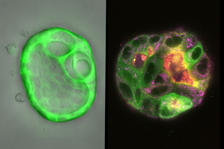
How Does The Cytoskeleton Transport Molecules?
VIDEO ON DEMAND - See how 3D cysts derived from MDCK cells help scientists understand how proteins are transported and recycled in tissues and the role of the cytoskeleton in this transport.
Loading...
 embryo, from sphere stage to somite stages.")
Studying Early Phase Development of Zebrafish Embryos
This video on demand focuses on combining widefield and confocal imaging to study the early-stage development of zebrafish embryos (Danio rerio), from oocyte to multicellular stage.
Loading...

How AR Helps in the Surgical Treatment of Moyamoya Disease
Moyamoya disease is a rare chronic occlusive cerebrovascular disorder characterized by progressive stenosis in the terminal portion of the internal carotid artery and an abnormal vascular network at…
Loading...

Multi-Color Caspase 3/7 Assays with Mica
Caspases are involved in apoptosis and can be utilized to determine if cells are undergoing this programmed cell death pathway in so-called caspase assays. These assays can be run by e.g. flow…
Loading...

How To Get Multi Label Experiment Data With Full Spatiotemporal Correlation
This video on demand focuses on the special challenges of live cell experiments. Our hosts Lynne Turnbull and Oliver Schlicker use the example of studying the mitochondrial activity of live cells.…
Loading...

Improving Ergonomics With a Dental Microscope
Here, we show you how Dr. David Blanc, a dental surgeon and ergonomics consultant, improves physical comfort using a dental microscope with ultra-low binoculars. Through optimized ergonomics, Dr.…
Loading...

Using GLOW800 AR in Radial Forearm Flap Free Phalloplasty
In this video, Chief Microsurgeon Professor Küntscher and his team perform a radial forearm free flap phalloplasty and use ICG fluorescence imaging to show the blood flow in the whole flap from the…
Loading...

Find Relevant Specimen Details from Overviews
Switch from searching image by image to seeing the full overview of samples quickly and identifying the important specimen details instantly with confocal microscopy. Use that knowledge to set up…
