
Science Lab
Science Lab
The knowledge portal of Leica Microsystems offers scientific research and teaching material on the subjects of microscopy. The content is designed to support beginners, experienced practitioners and scientists alike in their everyday work and experiments. Explore interactive tutorials and application notes, discover the basics of microscopy as well as high-end technologies – become part of the Science Lab community and share your expertise!
Filter articles
Tags
Story Type
Products
Loading...

Optimizing THUNDER Platform for High-Content Slide Scanning
With rising demand for full-tissue imaging and the need for FL signal quantitation in diverse biological specimens, the limits on HC imaging technology are tested, while user trainability and…
Loading...
Visualizing Protein Degradation and Aggregation in the Living Cell
Our guest speaker, Prof Dr Eric Reits, presents his work on neurodegenerative disorders. Reits’ group are experts on the subject of Huntington’s disease and work towards identifying leads for…
Loading...

Towards Advanced Use of Intraoperative OCT in Cataract Surgery
In this White Paper, Dr. Rachid Tahiri shares his personal experience with the Leica EnFocus intraoperative OCT, the valuable features supporting smooth surgery and how it allows him to minimize…
Loading...

How Clinical and Surgical Microscopes Support Otolaryngology
The number of ENT surgery procedures is increasing year after year. It is estimated that there will be over 21 million ENT procedures performed annually worldwide by 2022.
Dr. Duane Mol is the…
Loading...
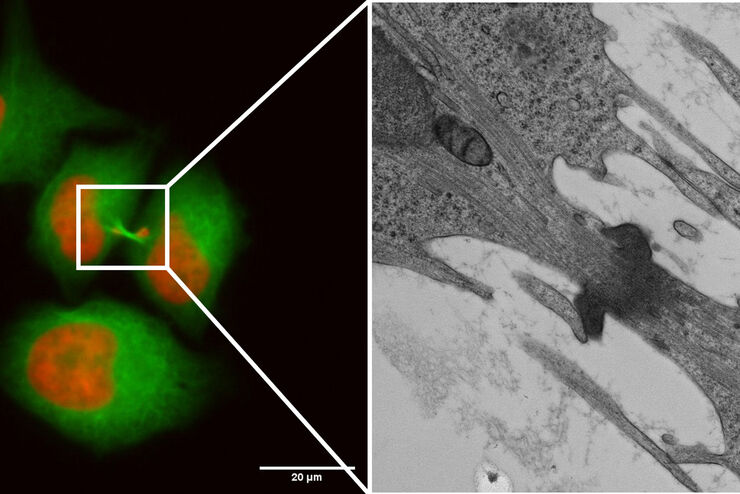
Capture life as it happens
With the Leica Nano Workflow, searching for the needle in the haystack is a thing of the past. Take advantage of correlative light and electron microscopy to identify directly the right cell at the…
Loading...
Life Beyond the Pixels: Deep Learning Methods for Single Cell Analysis
Our guest speaker Prof Dr Peter Horvath presents his work on single cell-based large-scale microscopy experiments. This novel targeting approach includes the use of machine learning models and…
Loading...

Introduction to 21 CFR Part 11 and Related Regulations
This article provides an overview of regulations and guidelines for electronic records (data entry, storage, signatures, and approvals) used in the USA (21 CFR Part 11), EU (GMP Annex 11), and China…
Loading...

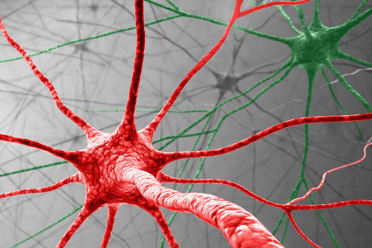
Physiology Image Gallery
Physiology is about the processes and functions within a living organism. Research in physiology focuses on the activities and functions of an organism’s organs, tissues, or cells, including the…
Loading...

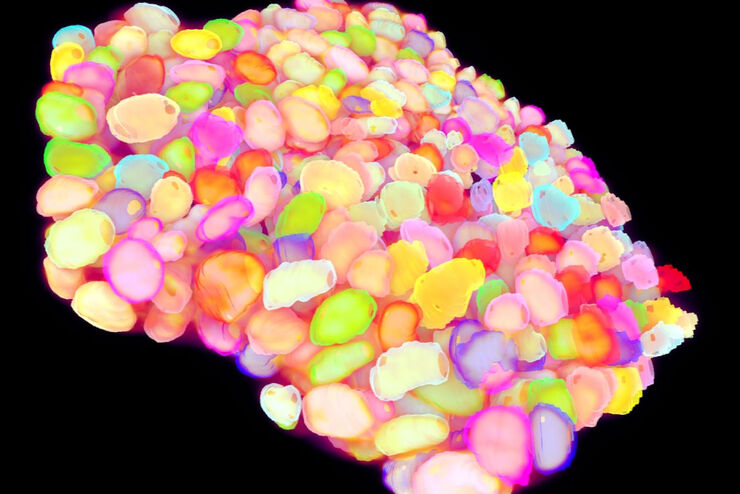
Live Cell Imaging Gallery
Live cell microscopy techniques are fundamental to get a better understanding of cellular and molecular function. Today, widefield microscopy is the most common technique used to visualize cell…
