
Science Lab
Science Lab
The knowledge portal of Leica Microsystems offers scientific research and teaching material on the subjects of microscopy. The content is designed to support beginners, experienced practitioners and scientists alike in their everyday work and experiments. Explore interactive tutorials and application notes, discover the basics of microscopy as well as high-end technologies – become part of the Science Lab community and share your expertise!
Filter articles
Tags
Story Type
Products
Loading...
, actin network (ATTO 647N), and nuclear pore basket (CF 680R).")
The Guide to STED Sample Preparation
This guide is intended to help users optimize sample preparation for stimulated emission depletion (STED) nanoscopy, specifically when using the STED microscope from Leica Microsystems. It gives an…
Loading...

How to Streamline Your Histology Workflows
Streamline your histology workflows. The unique Fluosync detection method embedded into Mica enables high-res RGB color imaging in one shot.
Loading...
 dataset, showing the biochemically distinct structures of a fresh, untreated apple slice.")
How to Prepare Samples for Stimulated Raman Scattering (SRS) imaging
Find here guidelines for how to prepare samples for stimulated Raman scattering (SRS), acquire images, analyze data, and develop suitable workflows. SRS spectroscopic imaging is also known as SRS…
Loading...

RPE65 Gene Therapy Subretinal Injection: Benefits of Intraoperative OCT
Discover how RPE65 gene therapy subretinal injection procedures in patients with Leber congenital amaurosis is supported by intraoperative Optical Coherence Tomography.
Loading...
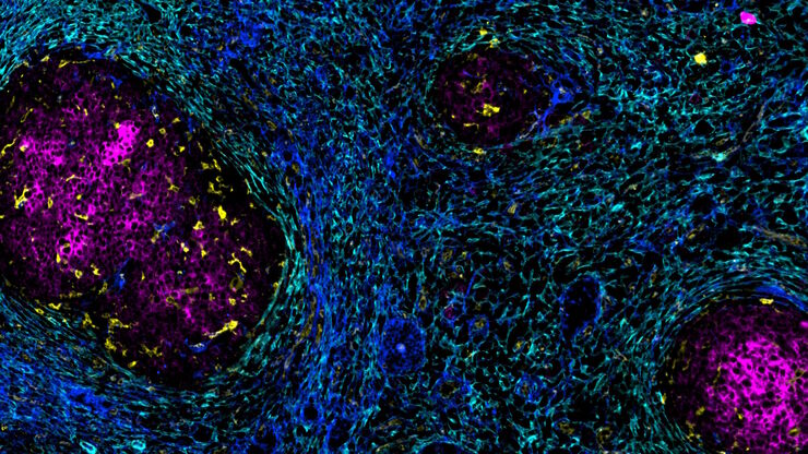
Accelerating Discovery for Multiplexed Imaging of Diverse Tissues
Explore IBEX: Open-source multiplexed imaging. Join the collaborative IBEX Imaging Community for optimized tissue processing, antibody selection, and human atlas construction.
Loading...

Key Factors for Efficient Cleanliness Analysis
An overview of the key factors necessary for technical cleanliness and efficient cleanliness analysis concerning automotive and electronics manufacturing and production is provided in this article.
Loading...

Technical Terms for Digital Microscope Cameras and Image Analysis
Learn more about the basic principles behind digital microscope camera technologies, how digital cameras work, and take advantage of a reference list of technical terms from this article.
Loading...
 and oblique (right) brightfield illumination using a Leica compound microscope. The defect on the wafer surface is clearly more visible with oblique illumination.")
Rapid Semiconductor Inspection with Microscope Contrast Methods
Semiconductor inspection during the production of patterned wafers and ICs (integrated circuits) is important for identifying and minimizing defects. To increase the efficiency of quality control in…
Loading...

Transforming Multiplexed 2D Data into Spatial Insights Guided by AI
Aivia 13 handles large 2D images and enables researchers to obtain deep insights into microenvironment surrounding their phenotypes with millions of detected objects and automatic clustering up to 30…
