
Science Lab
Science Lab
The knowledge portal of Leica Microsystems offers scientific research and teaching material on the subjects of microscopy. The content is designed to support beginners, experienced practitioners and scientists alike in their everyday work and experiments. Explore interactive tutorials and application notes, discover the basics of microscopy as well as high-end technologies – become part of the Science Lab community and share your expertise!
Filter articles
Tags
Story Type
Products
Loading...

Deep Visual Proteomics Provides Precise Spatial Proteomic Information
Despite the availability of imaging methods and mass spectroscopy for spatial proteomics, a key challenge that remains is correlating images with single-cell resolution to protein-abundance…
Loading...

What is Empty Magnification and How can Users Avoid it
The phenomenon of “empty magnification”, which can occur while using an optical, light, or digital microscope, and how it can be avoided is explained in this article. The performance of an optical…
Loading...

How to Study Gene Regulatory Networks in Embryonic Development
Join Dr. Andrea Boni by attending this on-demand webinar to explore how light-sheet microscopy revolutionizes developmental biology. This advanced imaging technique allows for high-speed, volumetric…
Loading...

Spatial Analysis of Neuroimmune Interactions in Alzheimer’s Disease
Alzheimer’s disease (AD) is a complex neurodegenerative disorder characterized by neurofibrillary tangles, β-amyloid plaques, and neuroinflammation. These dysfunctions trigger or are exacerbated by…
Loading...
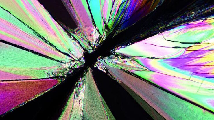
The Polarization Microscopy Principle
Polarization microscopy is routinely used in the material and earth sciences to identify materials and minerals on the basis of their characteristic refractive properties and colors. In biology,…
Loading...

A Guide to Spatial Biology
What is spatial biology, and how can researchers leverage its tools to meet the growing demands of biological questions in the post-omics era? This article provides a brief overview of spatial biology…
Loading...

An Introduction to Laser Microdissection
The heterogeneity of histological and biological specimens often requires isolation of specific single cells or cell groups from surrounding tissue before molecular biology analysis can be carried…
Loading...

Cutting-Edge Imaging Techniques for GPCR Signaling
With this webinar on-demand enhance your pharmacological research with our webinar on GPCR signaling and explore cutting-edge imaging techniques that aim to understand how GPCR signaling translates…
Loading...

Revealing Neuronal Migration’s Molecular Secrets
Different approaches can be used to investigate neuronal migration to their niche in the developing brain. In this webinar, experts from The University of Oxford present the microscopy tools and…
