
Science Lab
Science Lab
The knowledge portal of Leica Microsystems offers scientific research and teaching material on the subjects of microscopy. The content is designed to support beginners, experienced practitioners and scientists alike in their everyday work and experiments. Explore interactive tutorials and application notes, discover the basics of microscopy as well as high-end technologies – become part of the Science Lab community and share your expertise!
Filter articles
Tags
Story Type
Products
Loading...

A Guide to Darkfield Microscopes
A darkfield microscope offers a way to view the structures of many types of biological specimens in greater contrast without the need of stains.
Loading...
A Guide to Differential Interference Contrast (DIC)
A DIC microscope is a widefield microscopy which has a polarization filter and Wollaston prism between the light source and condenser lens and also between the objective lens and camera sensor or…
Loading...

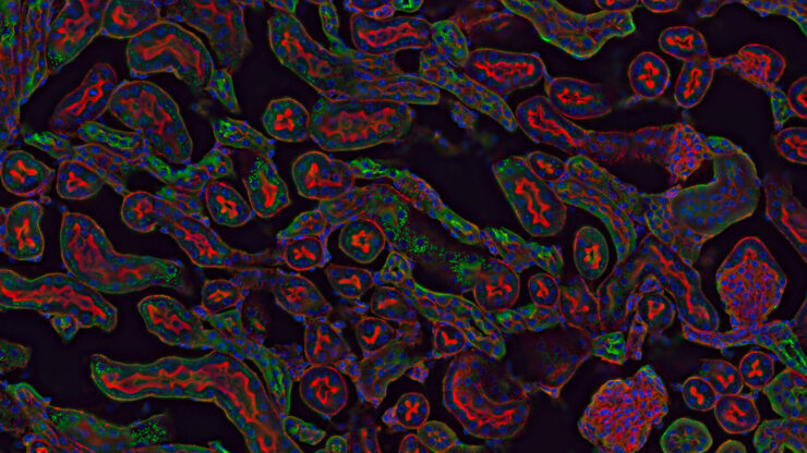
A Practical Guide to Virology Research
Leica solutions for imaging and sample preparation help you with the investigation of viral entry and fusion, genome integration, viral replication, assembly, and virus budding.
Loading...
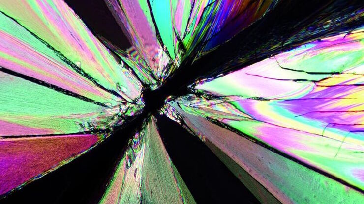
The Polarization Microscopy Principle
Polarization microscopy is routinely used in the material and earth sciences to identify materials and minerals on the basis of their characteristic refractive properties and colors. In biology,…
Loading...

Epi-Illumination Fluorescence and Reflection-Contrast Microscopy
This article discusses the development of epi-illumination and reflection contrast for fluorescence microscopy concerning life-science applications. Much was done by the Ploem research group…
Loading...

Differential Interference Contrast (DIC) Microscopy
This article demonstrates how differential interference contrast (DIC) can be actually better than brightfield illumination when using microscopy to image unstained biological specimens.
Loading...
 cells taken with phase contrast.")
Phase Contrast and Microscopy
This article explains phase contrast, an optical microscopy technique, which reveals fine details of unstained, transparent specimens that are difficult to see with common brightfield illumination.
Loading...
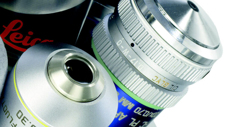
Immersion Objectives
How an immersion objective, which has a liquid medium between it and the specimen being observed, helps increase the numerical aperture and microscope resolution is explained in this article.
Loading...
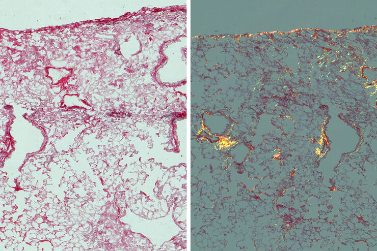
Studying Pulmonary Fibrosis
The results shown in this article demonstrate that fibrotic and non-fibrotic regions of collagen present in mouse lung tissue can be distinguished better with polarized light compared to brightfield.…
