
Science Lab
Science Lab
The knowledge portal of Leica Microsystems offers scientific research and teaching material on the subjects of microscopy. The content is designed to support beginners, experienced practitioners and scientists alike in their everyday work and experiments. Explore interactive tutorials and application notes, discover the basics of microscopy as well as high-end technologies – become part of the Science Lab community and share your expertise!
Filter articles
Tags
Story Type
Products
Loading...
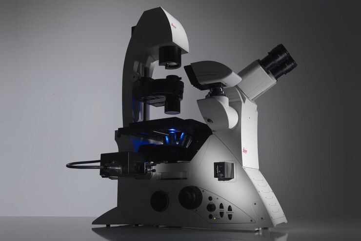
Factors to Consider When Selecting a Research Microscope
An optical microscope is often one of the central devices in a life-science research lab. It can be used for various applications which shed light on many scientific questions. Thereby the…
Loading...
The Fundamentals and History of Fluorescence and Quantum Dots
At some point in your research and science career, you will no doubt come across fluorescence microscopy. This ubiquitous technique has transformed the way in which microscopists can image, tag and…
Loading...

Koehler Illumination: A Brief History and a Practical Set Up in Five Easy Steps
In this article, we will look at the history of the technique of Koehler Illumination in addition to how to adjust the components in five easy steps.
Loading...
 illuminated with wide-band UV excitation. Note the tissue structure with blue autofluorescence. Right: Same tissue and same immunostaining with FITC label illuminated with epi-illumination using narrow")
Milestones in Incident Light Fluorescence Microscopy
Since the middle of the last century, fluorescence microscopy developed into a bio scientific tool with one of the biggest impacts on our understanding of life. Watching cells and proteins with the…
Loading...

Video Tutorial: How to Change the Bulb of a Fluorescence Lamp Housing
When applying fluorescence microscopy in biological applications, a lamp housing with mercury burner is the most common light source. This video tutorial shows how to change the bulb of a traditional…
Loading...

Video Tutorial: How to Align the Bulb of a Fluorescence Lamp Housing
The traditional light source for fluorescence excitation is a fluorescence lamp housing with mercury burner. A prerequisite for achieving bright and homogeneous excitation is the correct centering and…
Loading...

Fluorescent Proteins - From the Beginnings to the Nobel Prize
Fluorescent proteins are the fundament of recent fluorescence microscopy and its modern applications. Their discovery and consequent development was one of the most exciting innovations for life…
Loading...

Optical Contrast Methods
Optical contrast methods give the potential to easily examine living and colorless specimens. Different microscopic techniques aim to change phase shifts caused by the interaction of light with the…
Loading...

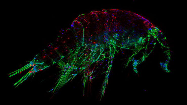
Fluorescence in Microscopy
Fluorescence microscopy is a special form of light microscopy. It uses the ability of fluorochromes to emit light after being excited with light of a certain wavelength. Proteins of interest can be…
