Contact Us
Science Lab

Science Lab

Science Lab

The knowledge portal of Leica Microsystems offers scientific research and teaching material on the subjects of microscopy. The content is designed to support beginners, experienced practitioners and scientists alike in their everyday work and experiments. Explore interactive tutorials and application notes, discover the basics of microscopy as well as high-end technologies – become part of the Science Lab community and share your expertise!

Primary Beam Splitting Devices for Confocal Microscopes

Current fluorescence microscopy employs incident illumination which requires separation of illumination and emission light. The classical device performing this separation is a color-dependent beam…
Pinhole diameter and diffraction pattern.

Pinhole Effect in Confocal Microscopes

When operating a confocal microscope, or when discussing features and parameters of such a device, we inescapably mention the pinhole and its diameter. This short introductory document is meant to…
Left: Tissue cells marked with an immunolabel (FITC) illuminated with wide-band UV excitation. Note the tissue structure with blue autofluorescence. Right: Same tissue and same immunostaining with FITC label illuminated with epi-illumination using narrow-band blue (490 nm) light. Note the increased image contrast (Ploem, 1967)

Milestones in Incident Light Fluorescence Microscopy

Since the middle of the last century, fluorescence microscopy developed into a bio scientific tool with one of the biggest impacts on our understanding of life. Watching cells and proteins with the…

BABB Clearing and Imaging for High Resolution Confocal Microscopy

Multipohoton microscopy experiment using Leica TCS SP8 MP and Leica 20x/0.95 NA BABB immersion objective. Understanding kidney microanatomy is key to detecting and identifying early events in kidney…
© wowomnom – Fotolia.com

From Light to Mind: Sensors and Measuring Techniques in Confocal Microscopy

This article outlines the most important sensors used in confocal microscopy. By confocal microscopy, we mean "True Confocal Scanning", i.e. the technique that illuminates and measures one single…
Acousto-optics, sketch

Acousto Optics in True Confocal Spectral Microscope Systems

Acousto-optical elements have successfully replaced planar filters in many positions. The white confocal, regarded as the fully spectrally tunable confocal microscope, was not possible without this…

Spectral Detection – How to Define the Spectral Bands that Collect Probe-specific Emission

To specifically collect emission from multiple probes, the light is first separated spatially and then passes through a device that defines a spectral band. Classically, this is a common glass-based…

Step by Step Guide for FRAP Experiments

Fluorescence Recovery After Photobleaching (FRAP) has been considered the most widely applied method for observing translational diffusion processes of macromolecules. The resulting information can be…

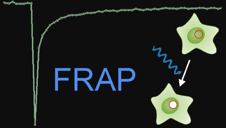
Fluorescence Recovery after Photobleaching (FRAP) and its Offspring

FRAP (Fluorescence recovery after photobleaching) can be used to study cellular protein dynamics: For visualization the protein of interest is fused to a fluorescent protein or a fluorescent dye. A…
Scroll to top