
Science Lab
Science Lab
The knowledge portal of Leica Microsystems offers scientific research and teaching material on the subjects of microscopy. The content is designed to support beginners, experienced practitioners and scientists alike in their everyday work and experiments. Explore interactive tutorials and application notes, discover the basics of microscopy as well as high-end technologies – become part of the Science Lab community and share your expertise!
Filter articles
Tags
Story Type
Products
Loading...

Accurately Analyze Fluorescent Widefield Images
The specificity of fluorescence microscopy allows researchers to accurately observe and analyze biological processes and structures quickly and easily, even when using thick or large samples. However,…
Loading...

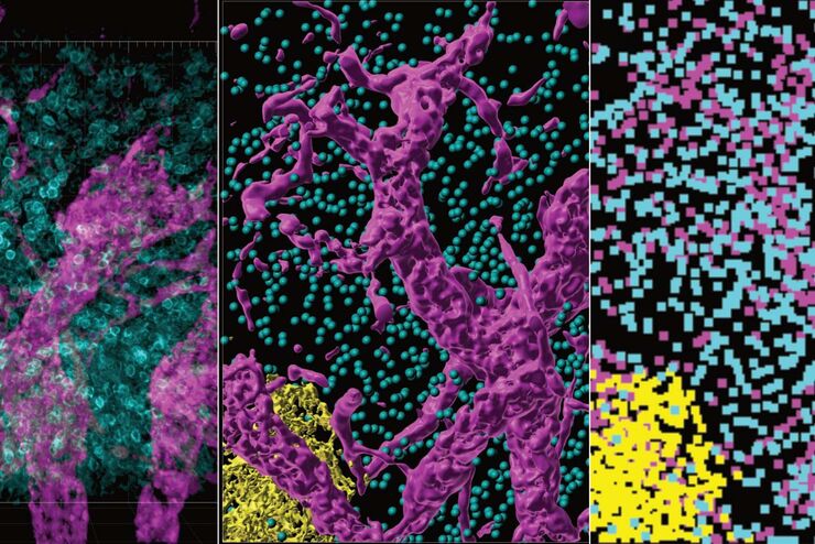
High-resolution 3D Imaging to Investigate Tissue Ageing
Award-winning researcher Dr. Anjali Kusumbe demonstrates age-related changes in vascular microenvironments through single-cell resolution 3D imaging of young and aged organs.
Loading...

Optimizing THUNDER Platform for High-Content Slide Scanning
With rising demand for full-tissue imaging and the need for FL signal quantitation in diverse biological specimens, the limits on HC imaging technology are tested, while user trainability and…
Loading...

Physiology Image Gallery
Physiology is about the processes and functions within a living organism. Research in physiology focuses on the activities and functions of an organism’s organs, tissues, or cells, including the…
Loading...

Developmental Biology Image Gallery
Developmental biology explores the development of complex organisms from the embryo to adulthood to understand in detail the origins of disease. This category of the gallery shows images about…
Loading...

The Power of Pairing Adaptive Deconvolution with Computational Clearing
Learn how deconvolution allows you to overcome losses in image resolution and contrast in widefield fluorescence microscopy due to the wave nature of light and the diffraction of light by optical…
Loading...
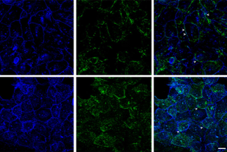
Improvement of Imaging Techniques to Understand Organelle Membrane Cell Dynamics
Understanding cell functions in normal and tumorous tissue is a key factor in advancing research of potential treatment strategies and understanding why some treatments might fail. Single-cell…
Loading...

Image Gallery: THUNDER Imager
To help you answer important scientific questions, THUNDER Imagers eliminate the out-of-focus blur that clouds the view of thick samples when using camera-based fluorescence microscopes. They achieve…
Loading...
From Organs to Tissues to Cells: Analyzing 3D Specimens with Widefield Microscopy
Obtaining high-quality data and images from thick 3D samples is challenging using traditional widefield microscopy because of the contribution of out-of-focus light. In this webinar, Falco Krüger…
