
Science Lab
Science Lab
The knowledge portal of Leica Microsystems offers scientific research and teaching material on the subjects of microscopy. The content is designed to support beginners, experienced practitioners and scientists alike in their everyday work and experiments. Explore interactive tutorials and application notes, discover the basics of microscopy as well as high-end technologies – become part of the Science Lab community and share your expertise!
Filter articles
Tags
Story Type
Products
Loading...

Introduction to Ultramicrotomy
When studying samples, to visualize their fine structure with nanometer scale resolution, most often electron microscopy is used. There are 2 types: scanning electron microscopy (SEM) which images the…
Loading...

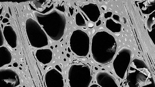
Expert Knowledge on High Pressure Freezing and Freeze Fracturing in the Cryo SEM Workflow
Get an insight in the working methods of the laboratory and learn about the advantages of Cryo SEM investigation in EM Sample Preparation. Find out how high pressure freezing, freeze fracturing and…
Loading...

Bridging Structure and Dynamics at the Nanoscale through Optogenetics and Electrical Stimulation
Nanoscale ultrastructural information is typically obtained by means of static imaging of a fixed and processed specimen. However, this is only a snapshot of one moment within a dynamic system in…
Loading...
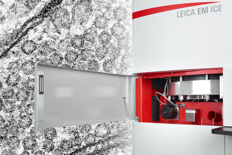
Expanding the Limits of Electron Microscopy Sample Preparation
Capturing the intricate changes in fine structure or in cell dynamics with conventional cryo solutions can be challenging sometimes. Leica Microsystems has developed a new cryo platform, the Leica EM…
Loading...

High Resolution Array Tomography with Automated Serial Sectioning
The optimization of high resolution, 3-dimensional (3D), sub-cellular structure analysis with array tomography using an automated serial sectioning solution, achieving a high section density on the…
Loading...

Macroscale to Nanoscale Pore Analysis of Shale and Carbonate Rocks
Physical porosity in rocks, like shale and carbonate, has a large effect on the their storage capacity. The pore geometries also affect their permeability. Imaging the visible pore space provides…
Loading...

Visualization of DNA Molecules
Precise low angle rotary shadowing with heavy metals (like platinum) can be used in transmission electron microscopy (TEM) to observe molecular details of objects previously absorbed on a thin, low…
Loading...
Practical Applications of Broad Ion Beam Milling
Mechanical polishing can be time consuming and frustrating. It can also introduce unwanted artifacts when preparing cross-sectioned samples for electron backscatter diffraction (EBSD) in the scanning…
Loading...

Bacteria Protocol - Critical Point Drying of E. coli for SEM
Application Note for Leica EM CPD300 - Critical point drying of E. coli with subsequent platinum / palladium coating and SEM analysis. Sample was inserted into a filter disc (Pore size: 16 - 40 μm)…
