
Science Lab
Science Lab
The knowledge portal of Leica Microsystems offers scientific research and teaching material on the subjects of microscopy. The content is designed to support beginners, experienced practitioners and scientists alike in their everyday work and experiments. Explore interactive tutorials and application notes, discover the basics of microscopy as well as high-end technologies – become part of the Science Lab community and share your expertise!
Filter articles
Tags
Story Type
Products
Loading...
")
Epoxy Resin Embedding of Animal and Human Tissues for Pathological Diagnosis and Research
Application Note for Leica EM AMW - The tissues were fixed in the modified Karnovsky fixative generally by immersion overnight (at minimum 4h at room temperature). Then pieces of approx. 1mm3 were cut…
Loading...

Brief Introduction to Freeze Substitution
Freeze-substitution is a process of dehydration, performed at temperatures low enough to avoid the formation of ice crystals and to circumvent the damaging effects observed after ambient-temperature…
Loading...

Brief Introduction to High-Pressure Freezing
Water is the most abundant cellular constituent and therefore important for preserving cellular ultra-structure. Currently the only way to fix cellular constituents without introducing significant…
Loading...
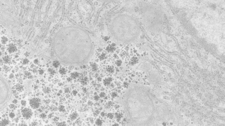
Perusing Alternatives for Automated Staining of TEM Thin Sections
Contrast in transmission electron microscopy (TEM) is mainly produced by electron scattering at the specimen: Structures that strongly scatter electrons are referred to as electron dense and appear as…
Loading...
An Introduction to CARS Microscopy
CARS overcomes the drawbacks of conventional staining methods by the intrinsic characteristics of the method. CARS does not require labeling because it is highly specific to molecular compounds which…
Loading...
")
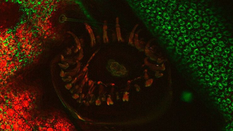
Mosaic Images
Confocal laser scanning microscopes are widely used to create highly resolved 3D images of cells, subcellular structures and even single molecules. Still, an increasing number of scientists are…
