
Science Lab
Science Lab
The knowledge portal of Leica Microsystems offers scientific research and teaching material on the subjects of microscopy. The content is designed to support beginners, experienced practitioners and scientists alike in their everyday work and experiments. Explore interactive tutorials and application notes, discover the basics of microscopy as well as high-end technologies – become part of the Science Lab community and share your expertise!
Filter articles
Tags
Story Type
Products
Loading...
, unsaturated lipids (magenta, 3050 cm-1), collagen (SHG, cyan). Sample courtesy of R. Rudolf, J Klicks, Hochschule Mannheim")
The Potential of Coherent Raman Scattering Microscopy at a Glance
Coherent Raman scattering microscopy (CRS) is a powerful approach for label-free, chemically specific imaging. It is based on the characteristic intrinsic vibrational contrast of molecules in the…
Loading...
Visualizing Protein Degradation and Aggregation in the Living Cell
Our guest speaker, Prof Dr Eric Reits, presents his work on neurodegenerative disorders. Reits’ group are experts on the subject of Huntington’s disease and work towards identifying leads for…
Loading...

Tissue Image Gallery
Visual analysis of animal and human tissues is critical to understand complex diseases such as cancer or neurodegeneration. From basic immunohistochemistry to intravital imaging, confocal microscopy…
Loading...
 and astrocytes (green) in a cortical spheroid derived from human induced pluripotent stem cells.")
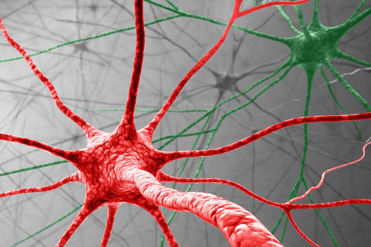
Neuroscience Images
Neuroscience commonly uses microscopy to study the nervous system’s function and understand neurodegenerative diseases.
Loading...

20 Years of Leica Laser Microdissection
Phenotype-genotype correlations are key for insight. From Eye to Insight is therefore fitting perfectly to Leica Microsystems and in particular to laser microdissection. Laser Microdissection, also…
Loading...

Chronic Inflammation Under the Microscope
In the course of chronic inflammation certain body areas are recurrently inflamed. This goes along with many human diseases. With the help of widefield light microscopy, the underlying processes can…
Loading...

CARS Microscopy: Imaging Characteristic Vibrational Contrast of Molecules
Coherent anti-Stokes Raman scattering (CARS) microscopy is a technique that generates images based on the vibrational signatures of molecules. This imaging methods does not require labeling, yet…
