
Science Lab
Science Lab
The knowledge portal of Leica Microsystems offers scientific research and teaching material on the subjects of microscopy. The content is designed to support beginners, experienced practitioners and scientists alike in their everyday work and experiments. Explore interactive tutorials and application notes, discover the basics of microscopy as well as high-end technologies – become part of the Science Lab community and share your expertise!
Filter articles
Tags
Story Type
Products
Loading...
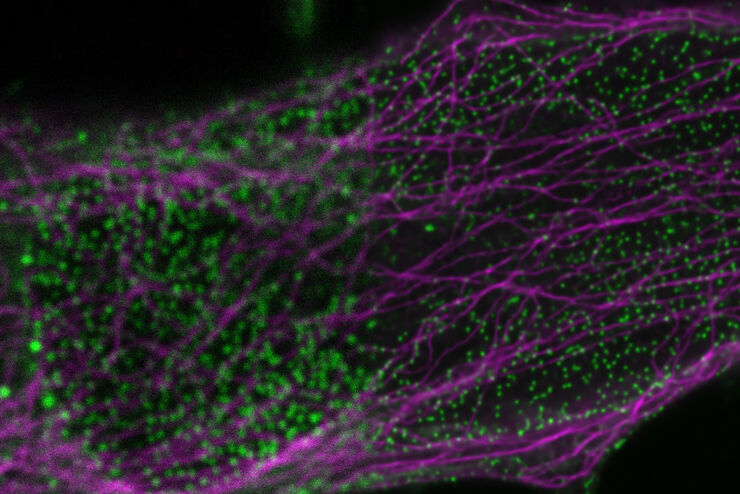
Fluorescence Lifetime-based Imaging Gallery
Confocal microscopy relies on the effective excitation of fluorescence probes and the efficient collection of photons emitted from the fluorescence process. One aspect of fluorescence is the emission…
Loading...
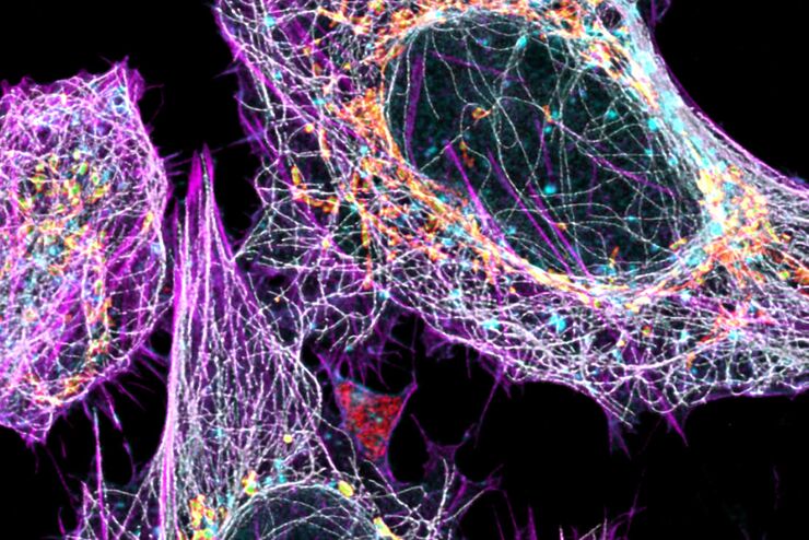
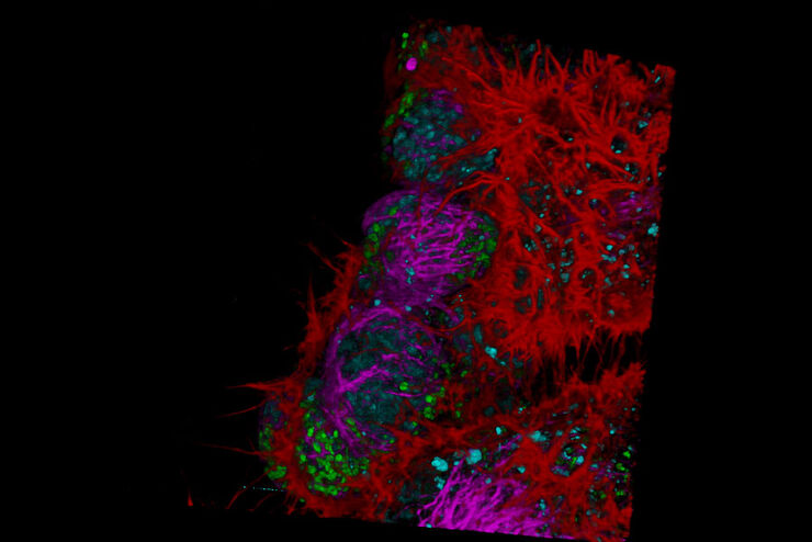
Multicolor Image Gallery
Fluorescence multicolor microscopy, which is one aspect of multiplex imaging, allows for the observation and analysis of multiple elements within the same sample – each tagged with a different…
Loading...
AI in Microscopy Webinar
We demonstrate residual channel attention networks for restoring and enhancing volumetric time-lapse (4D) fluorescence microscopy data.
Loading...

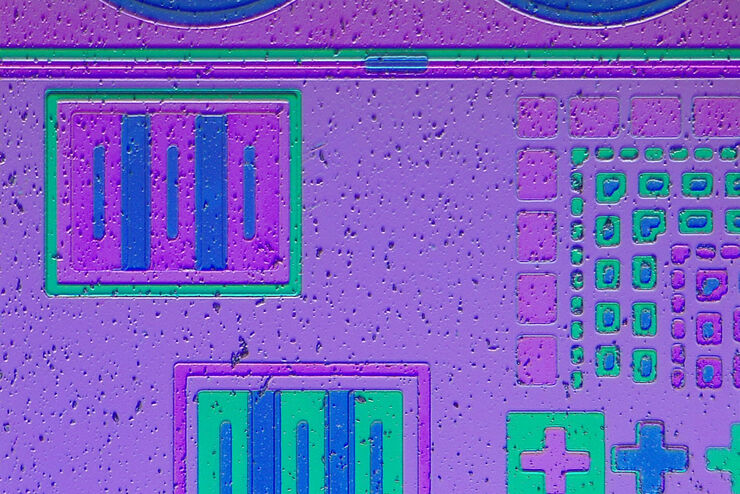
Topographic Analysis of Firing Pin Impressions on Cartridge Cases
The analysis of fired cartridges for primer cup morphology and flattening and firing pin impression (crater) depth using topographic data is discussed in this article. Topographical analysis of the…
Loading...

How does an Automated Rating Solution for Steel Inclusions Work?
The rating of non-metallic inclusions (NMIs) to determine steel quality is critical for many industrial applications. For an efficient and cost-effective steel quality evaluation, an automated NMI…
Loading...

How to Conduct Standard-Compliant Analysis of Non-Metallic Inclusions in Steel
This webinar will provide an overview of the significance of non-metallic inclusions in steel and outline the important global standards for rating the quality of steel and difficulties that arise in…
Loading...

An Introduction to Computational Clearing
Many software packages include background subtraction algorithms to enhance the contrast of features in the image by reducing background noise. The most common methods used to remove background noise…
Loading...

Challenges Faced When Manually Rating Non-Metallic Inclusions (NMIs) to Determine Steel Quality
Rapid, accurate, and reliable rating of non-metallic inclusions (NMIs) is instrumental for the determination of steel quality. This article describes the challenges that arise from manual NMI rating,…
Loading...
Industrial Microscopy: Digital imaging and the Leica DVM6
This webinar will discuss digital microscopy and Leica’s digital DVM6 microscope. We will navigate the difference between optical and digital magnification, explain the differences in optics, and…
