
Science Lab
Science Lab
The knowledge portal of Leica Microsystems offers scientific research and teaching material on the subjects of microscopy. The content is designed to support beginners, experienced practitioners and scientists alike in their everyday work and experiments. Explore interactive tutorials and application notes, discover the basics of microscopy as well as high-end technologies – become part of the Science Lab community and share your expertise!
Filter articles
Tags
Story Type
Products
Loading...

Uncover the Hidden Complexity of Colon Cancer with the Big Data Viewer
Colorectal cancer poses a significant health burden. While surgery is effective initially, some patients develop recurrent secondary disease with poor prognosis, necessitating advanced therapies like…
Loading...

Dive into Pancreatic Cancer Research with the Big Data Viewer
Pancreatic cancer, with a mortality rate near 40%, is challenging to treat due to its proximity to major organs. This story explores the complex biology of pancreatic ductal adenocarcinoma (PDAC),…
Loading...

Overcoming Challenges with Microscopy when Imaging Moving Zebrafish Larvae
Zebrafish is a valuable model organism with many beneficial traits. However, imaging a full organism poses challenges as it is not stationary. Here, this case study shows how zebrafish larvae can be…
Loading...

How to Automatically Obtain Fluorescent Cells of Interest in a Block-face
Block-face created by automatic trimming under fluorescence.
Mammalian cells of interest, stained with CellTrackerTM Green are visualized within the block-face using the UC Enuity equipped with the…
Loading...
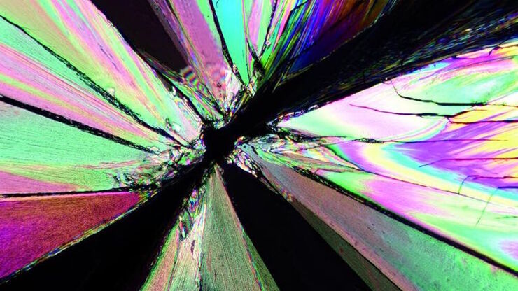
The Polarization Microscopy Principle
Polarization microscopy is routinely used in the material and earth sciences to identify materials and minerals on the basis of their characteristic refractive properties and colors. In biology,…
Loading...

An Introduction to Laser Microdissection
The heterogeneity of histological and biological specimens often requires isolation of specific single cells or cell groups from surrounding tissue before molecular biology analysis can be carried…
Loading...
 microdissected with a 10x objective (upper right). Inspection of the collection device (lower right).")
Molecular Biology Analysis facilitated with Laser Microdissection (LMD)
Extracting biomolecules, proteins, nucleic acids, lipids, and chromosomes, as well as extracting and manipulating cells and tissues with laser microdissection (LMD) enables insights to be gained into…
Loading...
.")
Neuron Isolation in Spatial Context with Laser Microdissection (LMD)
After Alzheimer’s disease, Parkinson’s is the second most common progressive neurodegenerative disease. Before the first symptoms manifest, up to 70% of dopamine-releasing neurons in the mid-brain…
Loading...
. With DIC users are able to visualize small height differences on the wafer surface more easily.")
6-Inch Wafer Inspection Microscope for Reliably Observing Small Height Differences
A 6-inch wafer inspection microscope with automated and reproducible DIC (differential interference contrast) imaging, no matter the skill level of users, is described in this article. Manufacturing…
