
Science Lab
Science Lab
The knowledge portal of Leica Microsystems offers scientific research and teaching material on the subjects of microscopy. The content is designed to support beginners, experienced practitioners and scientists alike in their everyday work and experiments. Explore interactive tutorials and application notes, discover the basics of microscopy as well as high-end technologies – become part of the Science Lab community and share your expertise!
Filter articles
Tags
Story Type
Products
Loading...
")
125 Years of Comparison Microscopy
To be able to optically compare two objects with scientific accuracy, it must be possible to view them at the same time. This is particularly true for comparing small objects that can only be…
Loading...

Acousto Optics in True Confocal Spectral Microscope Systems
Acousto-optical elements have successfully replaced planar filters in many positions. The white confocal, regarded as the fully spectrally tunable confocal microscope, was not possible without this…
Loading...
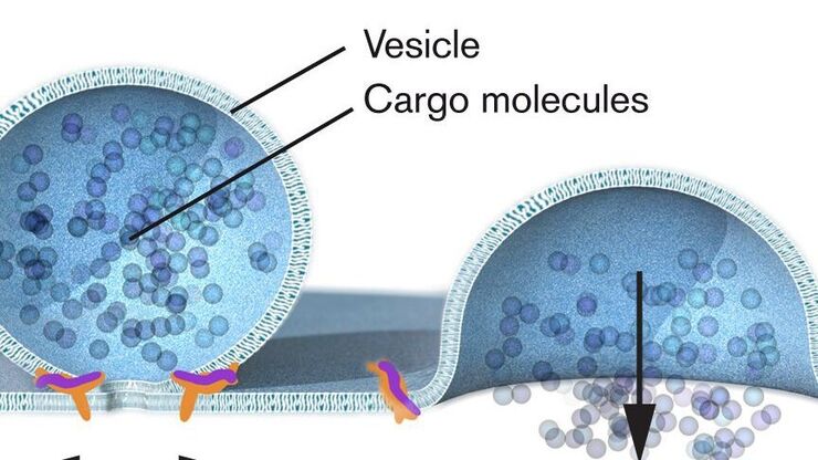
Nobel Prize 2013 in Physiology or Medicine for Discoveries of the Machinery Regulating Vesicle Traffic
On October 7th 2013, The Nobel Assembly at Karolinska Institutet has decided to award The Nobel Prize in Physiology or Medicine 2012 jointly to James E. Rothman, Randy W. Schekman and Thomas C. Südhof…
Loading...

Thermodynamic Considerations Regarding the LN2 in a High Pressure Freezer
Employing liquid nitrogen (LN2) as a coolant in the complex process of high pressure freezing raises certain considerations regarding phase transition not only of the liquid sample to be frozen but…
Loading...
Carbon Thickness Evaluation in Electron Microscopy
The coating layers applied and used for electron microscopy imaging are commonly controlled and measured by quartz crystals. These crystals oscillate with a certain frequency (around 6 megahertz when…
Loading...
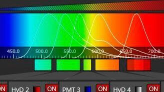
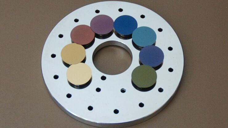
Spectral Detection – How to Define the Spectral Bands that Collect Probe-specific Emission
To specifically collect emission from multiple probes, the light is first separated spatially and then passes through a device that defines a spectral band. Classically, this is a common glass-based…
Loading...

Every Clue Counts – Forensics Inconceivable Without Microscopy
There is no crime without clues. They may be obvious, like a cartridge case at the scene of the crime or clear signs of crowbar damage on a door. But sometimes, clues are microscopically small.…
Loading...
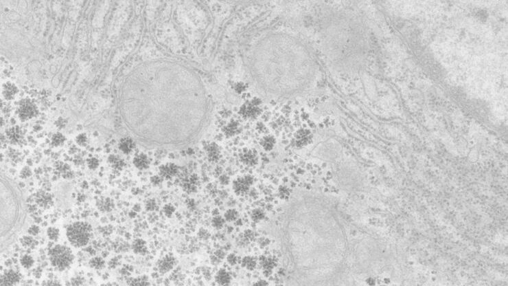
Perusing Alternatives for Automated Staining of TEM Thin Sections
Contrast in transmission electron microscopy (TEM) is mainly produced by electron scattering at the specimen: Structures that strongly scatter electrons are referred to as electron dense and appear as…
Loading...

50 Years of Image Analysis
Modern image analysis systems perform highly sophisticated image processing functions on images from an automated microscope and digital camera. 50 years ago, the first image analysis system was…
