
Science Lab
Science Lab
The knowledge portal of Leica Microsystems offers scientific research and teaching material on the subjects of microscopy. The content is designed to support beginners, experienced practitioners and scientists alike in their everyday work and experiments. Explore interactive tutorials and application notes, discover the basics of microscopy as well as high-end technologies – become part of the Science Lab community and share your expertise!
Filter articles
Tags
Story Type
Products
Loading...
 taken with a ring light (RL) and near vertical illumination (NVI).")
Microscope Illumination for Industrial Applications
Inspection microscope users can obtain information from this article which helps them choose the optimal microscope illumination or lighting system for inspection of parts or components.
Loading...
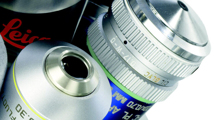
Immersion Objectives
How an immersion objective, which has a liquid medium between it and the specimen being observed, helps increase the numerical aperture and microscope resolution is explained in this article.
Loading...
 of an image of two points where the distance between them corresponds to the Rayleigh criterion.")
Microscope Resolution: Concepts, Factors and Calculation
This article explains in simple terms microscope resolution concepts, like the Airy disc, Abbe diffraction limit, Rayleigh criterion, and full width half max (FWHM). It also discusses the history.
Loading...
, SPY-Actin (cyan), and SiR-Tubulin (magenta). Instant Computational Clearing (ICC) was applied.")
How to Perform Dynamic Multicolor Time-Lapse Imaging
Live-cell imaging sheds light on diverse cellular events. As many of these events have fast dynamics, the microscope imaging system must be fast enough to record every detail. One major advantage of…
Loading...

Multiplexing through Spectral Separation of 11 Colors
Fluorescence microscopy is a fundamental tool for life science research that has evolved and matured together with the development of multicolor labeling strategies in cells tissues and model…
Loading...

RNA Quality after Different Tissue Sample Preparation
The influence of sample preparation and ultraviolet (UV) laser microdissection (UV LMD) on the quality of RNA from murine-brain tissue cryo-sections is described in this article. To obtain good…
Loading...

H&E Staining in Microscopy
If we consider the role of microscopy in pathologists’ daily routines, we often think of the diagnosis. While microscopes indeed play a crucial role at this stage of the pathology lab workflow, they…
Loading...

Cleanliness Analysis for Particulate Contamination
Devices, products, and their components fabricated in many industries can be quite sensitive to contamination and, as a result, have stringent requirements for technical cleanliness. Measurement…
Loading...

Efficient Particle Counting and Analysis
This report discusses particle counting and analysis using optical microscopy for cleanliness of parts and components. Particle counting and analysis is a critical part of quality assurance in the…
