
Science Lab
Science Lab
The knowledge portal of Leica Microsystems offers scientific research and teaching material on the subjects of microscopy. The content is designed to support beginners, experienced practitioners and scientists alike in their everyday work and experiments. Explore interactive tutorials and application notes, discover the basics of microscopy as well as high-end technologies – become part of the Science Lab community and share your expertise!
Filter articles
Tags
Story Type
Products
Loading...

A Practical Guide to Virology Research
Leica solutions for imaging and sample preparation help you with the investigation of viral entry and fusion, genome integration, viral replication, assembly, and virus budding.
Loading...
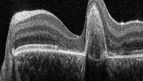
A Guide to OCT
Leica Optical Coherence Tomography (OCT) systems support ophthalmologists, ophthalmic surgeons, and researchers with easy-to-use, high-quality imaging technology.
Loading...

A Guide to Zebrafish Research
For the best result during screening, sorting, manipulation, and imaging you need to see details and structures to make the right decisions for your next steps in research.
Known for outstanding…
Loading...

A Guide to Cryo-Electron Tomography
Cryo-electron tomography (CryoET) is used to resolve biomolecules within their cellular environment down to an unprecedented resolution below one nanometer.
Loading...

A Guide to Spatial Biology
What is spatial biology, and how can researchers leverage its tools to meet the growing demands of biological questions in the post-omics era? This article provides a brief overview of spatial biology…
Loading...

Technical Terms for Digital Microscope Cameras and Image Analysis
Learn more about the basic principles behind digital microscope camera technologies, how digital cameras work, and take advantage of a reference list of technical terms from this article.
Loading...

Key Factors to Consider When Selecting a Stereo Microscope
This article explains key factors that help users determine which stereo microscope solution can best meet their needs, depending on the application.
Loading...
 in 14 x 18 tiles. Lifetime gives an additional contrast that allows to differentiate different structures in histological stainings.")
A Guide to Fluorescence Lifetime Imaging Microscopy (FLIM)
The fluorescence lifetime is a measure of how long a fluorophore remains on average in its excited state before returning to the ground state by emitting a fluorescence photon.
