
Science Lab
Science Lab
The knowledge portal of Leica Microsystems offers scientific research and teaching material on the subjects of microscopy. The content is designed to support beginners, experienced practitioners and scientists alike in their everyday work and experiments. Explore interactive tutorials and application notes, discover the basics of microscopy as well as high-end technologies – become part of the Science Lab community and share your expertise!
Filter articles
Tags
Story Type
Products
Loading...
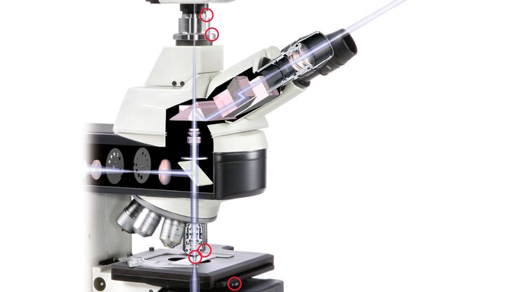
How to Clean Microscope Optics
Clean microscope optics are essential for obtaining good microscope images. If they are dirty, the microscope should be cleaned to avoid a loss of quality. If you decide to do this yourself, you…
Loading...
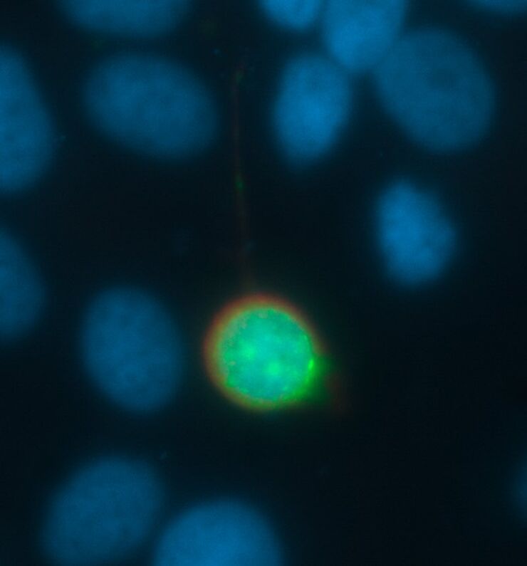
Image Processing for Widefield Microscopy
Fluorescence microscopy is a modern and steadily evolving tool to bring light to current cell biological questions. With the help of fluorescent proteins or dyes it is possible to make discrete…
Loading...

The Principles of White Light Laser Confocal Microscopy
The perfect light source for confocal microscopes in biomedical applications has sufficient intensity, tunable color and is pulsed for use in lifetime fluorescence. Furthermore, it should offer means…
Loading...
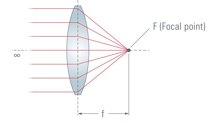
Optical Microscopes – Some Basics
The optical microscope has been a standard tool in life science as well as material science for more than one and a half centuries now. To use this tool economically and effectively, it helps a lot to…
Loading...
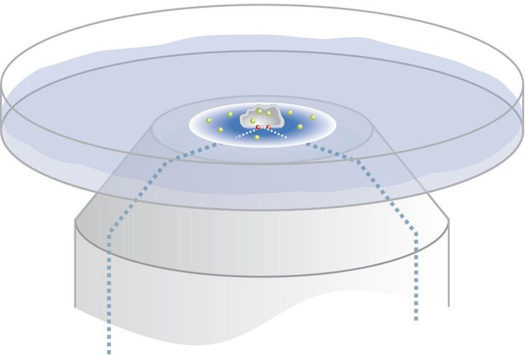
Controlling the TIRF Penetration Depth is Mandatory for Reproducible Results
The main feature of total internal reflection fluorescence (TIRF) microscopy is the employment of an evanescent wave for the excitation of fluorophores instead of using direct light. A property of the…
Loading...
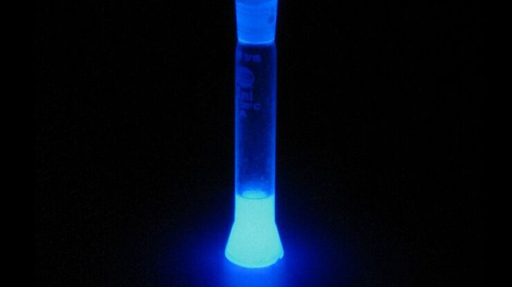
Basic Principles of Luminescence
There are a lot of light-emitting processes occurring in nature. Luminescence is an umbrella term for those kinds of events where light emission is not the result of high temperatures. This article…
Loading...

Introduction to Live-Cell Imaging
The understanding of complex and fast cellular dynamics is an important step to get insight into biological processes. Therefore, today’s life science research more and more demands studying…
Loading...
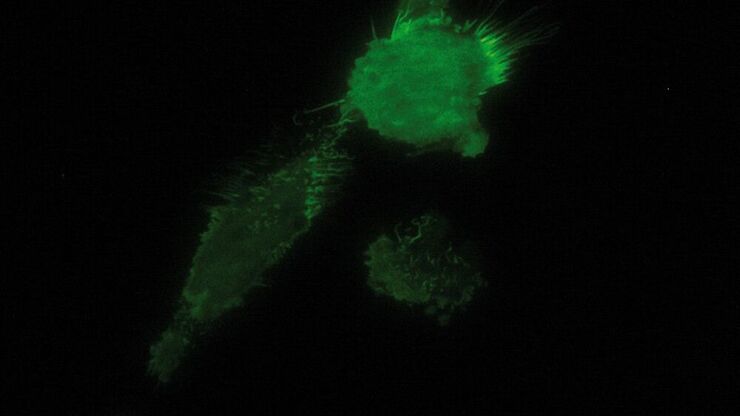
Total Internal Reflection Fluorescence (TIRF) Microscopy
Total internal reflection fluorescence (TIRF) is a special technique in fluorescence microscopy developed by Daniel Axelrod at the University of Michigan, Ann Arbor in the early 1980s. TIRF microscopy…
Loading...
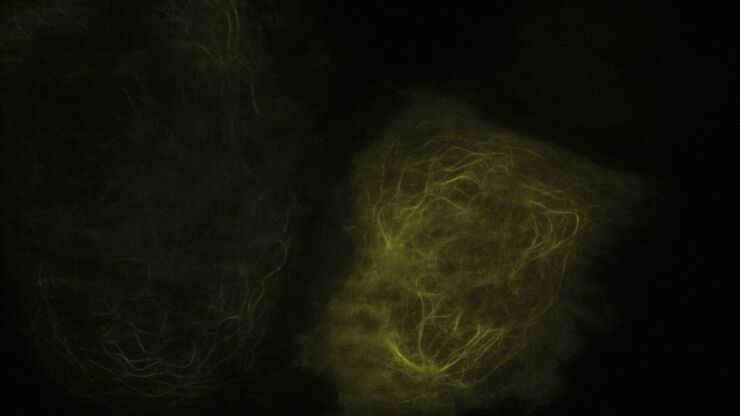
Applications of TIRF Microscopy in Life Science Research
The special feature of TIRF microscopy is the employment of an evanescent field for fluorophore excitation. Unlike standard widefield fluorescence illumination procedures with arc lamps, LEDs or…
