
Science Lab
Science Lab
The knowledge portal of Leica Microsystems offers scientific research and teaching material on the subjects of microscopy. The content is designed to support beginners, experienced practitioners and scientists alike in their everyday work and experiments. Explore interactive tutorials and application notes, discover the basics of microscopy as well as high-end technologies – become part of the Science Lab community and share your expertise!
Filter articles
Tags
Story Type
Products
Loading...

Considerations for Multiplex Live Cell Imaging
Simultaneous multicolor imaging for successful experiments: Live-cell imaging experiments are key to understand dynamic processes. They allow us to visually record cells in their living state, without…
Loading...

Imaging of Anti-Cancer Drug Uptake in Spheroids using DLS
Spheroid 3D cell culture models mimic the physiology and functions of living tissues making them a useful tool to study tumor morphology and screen anti-cancer drugs. The drug AZD2014 is a recognized…
Loading...

Top Challenges for Visual Inspection
This article discusses the challenges encountered when performing visual inspection and rework using a microscope. Using the right type of microscope and optical setup is paramount in order to…
Loading...
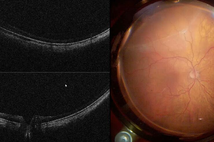
Intraoperative OCT in Retinal Procedures
In their white paper the surgical team at Retina Clinic in São Paulo provide insight into cases of complex retinal surgery in which OCT has provided helpful additional information which has on…
Loading...

How to Improve Live Cell Imaging with Coral Life
For live-cell CLEM applications, light microscopy imaging is a critical step for identifying the right cell in the right state at the right time. In this article, Leica experts share their insights on…
Loading...

How to Keep Your Samples Under Physiological Conditions
The Coral Life workflow combines dynamic data with the best possible sample fixation by high pressure freezing. However, good sample preservation won’t help if your cells are stressed by temperature…
Loading...

Towards Advanced Use of Intraoperative OCT in Cataract Surgery
In this White Paper, Dr. Rachid Tahiri shares his personal experience with the Leica EnFocus intraoperative OCT, the valuable features supporting smooth surgery and how it allows him to minimize…
Loading...
 and astrocytes (green) in a cortical spheroid derived from human induced pluripotent stem cells.")
Download The Guide to Live Cell Imaging
In life science research, live cell imaging is an indispensable tool to visualize cells in a state as in vivo as possible. This E-book reviews a wide range of important considerations to take to…
Loading...

Why is Manual Visual Inspection of Medical Devices so Challenging?
This article discusses how manual visual inspection, which is prevalent in the medical device industry, can lead to inconsistent results. It also addresses the challenges quality managers and…
