
Science Lab
Science Lab
Learn. Share. Contribute. The knowledge portal of Leica Microsystems. Find scientific research and teaching material on the subject of microscopy. The portal supports beginners, experienced practitioners and scientists alike in their everyday work and experiments. Explore interactive tutorials and application notes, discover the basics of microscopy as well as high-end technologies. Become part of the Science Lab community and share your expertise.
Filter articles
Tags
Story Type
Products
Loading...

From Bench to Beam: A Complete Correlative Cryo Light Microscopy Workflow
In the webinar entitled "A Multimodal Vitreous Crusade, a Cryo Correlative Workflow from Bench to Beam" a team of experts discusses the exciting world of correlative workflows for structural biology…
Loading...
Harnessing Microfluidics to Maintain Cell Health During Live-Cell Imaging
VIDEO ON DEMAND - In this webinar on-demand, we will use microfluidics to explore the effect of shear stress on cell morphology, examine the effect of nutrient replenishment on cellular growth during…
Loading...
 and CellEvent™ (yellow).")
Following Multiple Events during Staurosporine Apoptosis
In this video on demand, we show how adding additional markers to an apoptosis kit can markedly increase the amount of information a researcher can obtain from the same experiment. The simultaneous…
Loading...
, the cis-golgi matrix protein GM130 (AF488, green), and the trans-golgi network membrane protein TGN46 (AF647, red).")
Golgi Organizational Changes in Response to Cell Stress
In this video on demand, our special guest George Galea from EMBL Heidelberg will look at HeLa Kyoto cells treated with various chemotherapeutic agents to investigate their effect on the Golgi complex…
Loading...

Imaging of Cardiac Tissue Regeneration in Zebrafish
Learn how to image cardiac tissue regeneration in zebrafish focusing on cell proliferation and response during recovery with Laura Peces-Barba Castaño from the Max Planck Institute.
Loading...
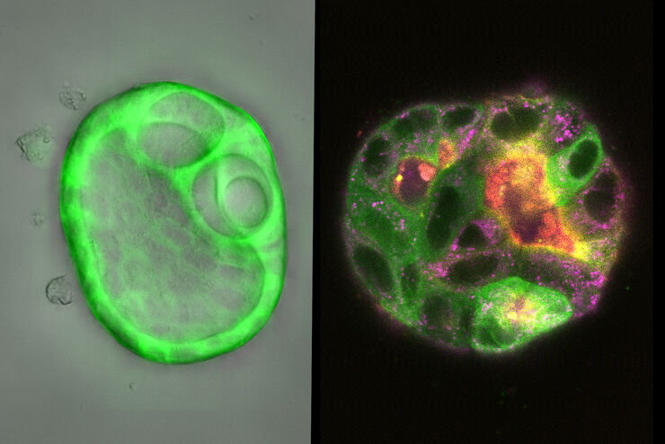
How Does The Cytoskeleton Transport Molecules?
VIDEO ON DEMAND - See how 3D cysts derived from MDCK cells help scientists understand how proteins are transported and recycled in tissues and the role of the cytoskeleton in this transport.
Loading...
 embryo, from sphere stage to somite stages.")
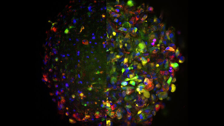
Studying Early Phase Development of Zebrafish Embryos
This video on demand focuses on combining widefield and confocal imaging to study the early-stage development of zebrafish embryos (Danio rerio), from oocyte to multicellular stage.
Loading...

How To Get Multi Label Experiment Data With Full Spatiotemporal Correlation
This video on demand focuses on the special challenges of live cell experiments. Our hosts Lynne Turnbull and Oliver Schlicker use the example of studying the mitochondrial activity of live cells.…
Loading...

How to Target Fluorescent Structures in 3D for Cryo-FIB Milling
This article describes the major steps of the cryo-electron tomography workflow including super-resolution cryo-confocal microscopy. We describe how subcellular structures can be precisely located in…
