
Science Lab
Science Lab
Learn. Share. Contribute. The knowledge portal of Leica Microsystems. Find scientific research and teaching material on the subject of microscopy. The portal supports beginners, experienced practitioners and scientists alike in their everyday work and experiments. Explore interactive tutorials and application notes, discover the basics of microscopy as well as high-end technologies. Become part of the Science Lab community and share your expertise.
Filter articles
Tags
Story Type
Products
Loading...

Accelerating Neuron Image Analysis with Automation
The ability to examine complex neural processes relies on the accurate reconstruction of neuronal networks at scale. Most data extraction methods in neuroscience research are time-consuming and…
Loading...

How to Improve Live Cell Imaging with Coral Life
For live-cell CLEM applications, light microscopy imaging is a critical step for identifying the right cell in the right state at the right time. In this article, Leica experts share their insights on…
Loading...
Fluorescence Lifetime-based Imaging Gallery
Confocal microscopy relies on the effective excitation of fluorescence probes and the efficient collection of photons emitted from the fluorescence process. One aspect of fluorescence is the emission…
Loading...

Optimizing THUNDER Platform for High-Content Slide Scanning
With rising demand for full-tissue imaging and the need for FL signal quantitation in diverse biological specimens, the limits on HC imaging technology are tested, while user trainability and…
Loading...

Physiology Image Gallery
Physiology is about the processes and functions within a living organism. Research in physiology focuses on the activities and functions of an organism’s organs, tissues, or cells, including the…
Loading...

Live Cell Imaging Gallery
Live cell microscopy techniques are fundamental to get a better understanding of cellular and molecular function. Today, widefield microscopy is the most common technique used to visualize cell…
Loading...

Tissue Image Gallery
Visual analysis of animal and human tissues is critical to understand complex diseases such as cancer or neurodegeneration. From basic immunohistochemistry to intravital imaging, confocal microscopy…
Loading...
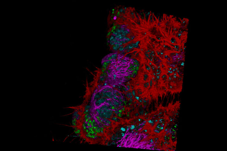
 and astrocytes (green) in a cortical spheroid derived from human induced pluripotent stem cells.")
Neuroscience Images
Neuroscience commonly uses microscopy to study the nervous system’s function and understand neurodegenerative diseases.
Loading...

Cancer Research Image Gallery
Fluorescence microscopy allows the study of changes occurring in tissue and cells during cancer development and progression. Techniques such as live cell imaging are critical to understand cancer…
