
Science Lab
Science Lab
Learn. Share. Contribute. The knowledge portal of Leica Microsystems. Find scientific research and teaching material on the subject of microscopy. The portal supports beginners, experienced practitioners and scientists alike in their everyday work and experiments. Explore interactive tutorials and application notes, discover the basics of microscopy as well as high-end technologies. Become part of the Science Lab community and share your expertise.
Filter articles
Tags
Story Type
Products
Loading...

Volume EM and AI Image Analysis
The article outlines a detailed workflow for studying biological tissues in three dimensions using volume-scanning electron microscopy (volume-SEM) combined with AI-assisted image analysis. The focus…
Loading...
Mastering Polymer Sectioning with Helmut Gnaegi
When it comes to ultramicrotomy, few names carry the weight of Helmut Gnaegi. As co-founder of Diatome, a global leader in diamond knife technology, Helmut has spent decades refining the art and…
Loading...
.")
How Fluorescence Guides Sectioning of Resin-embedded EM Samples
Electron microscopes, including transmission electron microscopes (TEM) and scanning electron microscopes (SEM), are widely utilized to gain detailed structural information about biological samples or…
Loading...

How to Save Time and Samples by Automated Ultramicrotomy
This article describes how 3D micro-CT data of a resin-embedded electron microscopy sample can be used to trim the specimen down to a defined target plane prior to sectioning. The interactive and…
Loading...
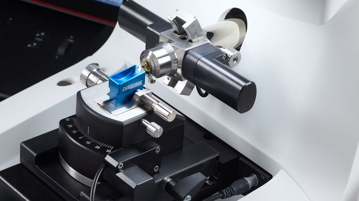
Essential Guide to Ultramicrotomy
When studying samples, to visualize their fine structure with nanometer scale resolution, most often electron microscopy is used. There are 2 types: scanning electron microscopy (SEM) which images the…
Loading...

How to Automatically Obtain Fluorescent Cells of Interest in a Block-face
Block-face created by automatic trimming under fluorescence.
Mammalian cells of interest, stained with CellTrackerTM Green are visualized within the block-face using the UC Enuity equipped with the…
Loading...

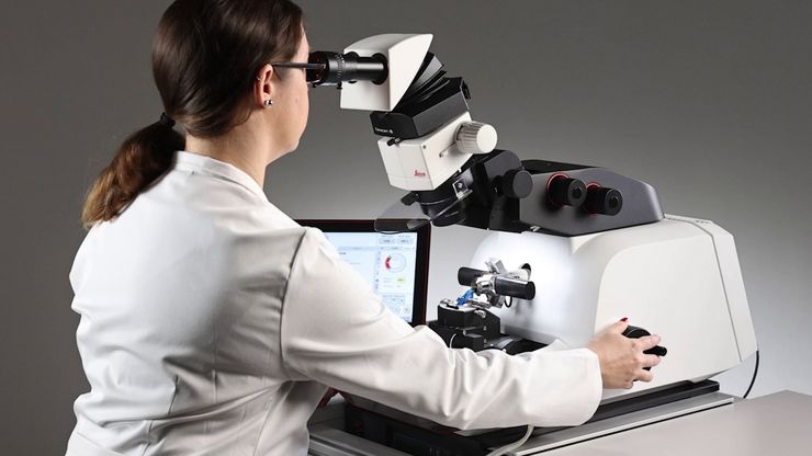
Improve Your Ultramicrotomy Workflow with Automated Sectioning
Discover advanced digital ultramicrotomy tools for fast and accurate automated sectioning. Learn about autoalignment, and efficient sample trimming leveraging 3D µCT data. See application examples…
Loading...

Automatic Alignment of Sample and Knife for High Sectioning Quality
Automatic alignment of sample and knife on the ultramicrotome UC Enuity, enabling even untrained users to create ultrathin sections with reduced risk of losing precious sections.
Loading...

High Quality Sectioning in Ultramicrotomy
Discover the significance of achieving high-quality uniform sections with ultramicrotomy for precise imaging in electron microscopy.
