
Science Lab
Science Lab
Learn. Share. Contribute. The knowledge portal of Leica Microsystems. Find scientific research and teaching material on the subject of microscopy. The portal supports beginners, experienced practitioners and scientists alike in their everyday work and experiments. Explore interactive tutorials and application notes, discover the basics of microscopy as well as high-end technologies. Become part of the Science Lab community and share your expertise.
Filter articles
Tags
Story Type
Products
Loading...
 observed with an Ivesta 3 stereo microscope during fly pushing (sorting of the flies). The scale bar length is 1 mm. Image courtesy of M. Benton, EMBL, Heidelberg, Germany.")
A Guide to Using Microscopy for Drosophila (Fruit Fly) Research
The fruit fly, typically Drosophila melanogaster, has been used as a model organism for over a century. One reason is that many disease-related genes are shared between Drosophila and humans. It is…
Loading...

A Guide to Neuroscience Research
Neuroscience often requires investigating challenging specimens to better understand the nervous system and disorders. Leica microscopes helps neuroscientists obtain insights into neuronal functions.
Loading...

Improving Zebrafish-Embryo Screening with Fast, High-Contrast Imaging
Discover from this article how screening of transgenic zebrafish embryos is boosted with high-speed, high-contrast imaging using the DM6 B microscope, ensuring accurate targeting for developmental…
Loading...

Unlocking the Secrets of Organoid Models in Biomedical Research
Get ready to delve deeper into the world of organoids and 3D models, which are essential tools for advancing our understanding of human health. Navigating these complex structures and obtaining clear…
Loading...

A Guide to Polarized Light Microscopy
Polarized light microscopy (POL) enhances contrast in birefringent materials and is used in geology, biology, and materials science to study minerals, crystals, fibers, and plant cell walls.
Loading...
 which was imaged with extended depth of field (EDOF) using digital microscopy.")
Depth of Field in Microscope Images
For microscopy imaging, depth of field is an important parameter when needing sharp images of sample areas with structures having significant changes in depth. In practice, depth of field is…
Loading...

Deep Visual Proteomics Provides Precise Spatial Proteomic Information
Despite the availability of imaging methods and mass spectroscopy for spatial proteomics, a key challenge that remains is correlating images with single-cell resolution to protein-abundance…
Loading...

What is Empty Magnification and How can Users Avoid it
The phenomenon of “empty magnification”, which can occur while using an optical, light, or digital microscope, and how it can be avoided is explained in this article. The performance of an optical…
Loading...
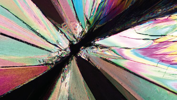
The Polarization Microscopy Principle
Polarization microscopy is routinely used in the material and earth sciences to identify materials and minerals on the basis of their characteristic refractive properties and colors. In biology,…
