Webinars
Take a look at all our upcoming congresses, exhibitions, webinars, and workshops and join us at one of our next events!
26
Feb
2025
Designing the Future: Novel Stem Cell Culture and RNA Tech
United Kingdom
•
Webinar
Filter articles
Tags
Products
Loading...
![Transfection using the Uncommon Bio reprogramming system. Image acquired using the THUNDER Imager 3D Cell Culture with THUNDER Large Volume Computational Clearing (LVCC) applied. Image courtesy of Samuel East, Uncommon Bio. Transfection using the Uncommon Bio reprogramming system. Image acquired using the THUNDER Imager 3D Cell Culture with THUNDER Large Volume Computational Clearing (LVCC) applied. Image courtesy of Samuel East, Uncommon Bio.](/fileadmin/_processed_/a/8/csm_Transfection_using_Uncommon_Bio_reprogramming_system_07fe8407a3.jpg)
Designing the Future with Stem Cell and RNA Technology
Visionary biotech start-up Uncommon Bio is tackling one of the world’s biggest health challenges: food sustainability. In this webinar, Stem Cell Scientist Samuel East will show how they use RNA…
Loading...
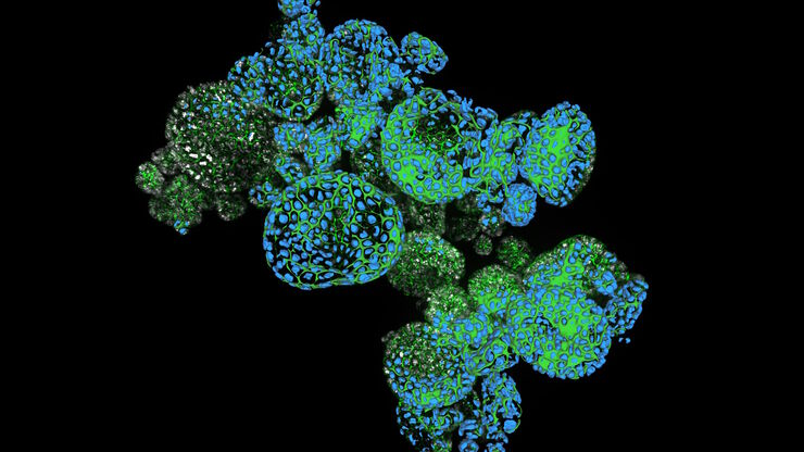
![Dapi – Nucleus, GFP – Plasma Membrane, Thickness 100µm, 63x objektive, 469 Z planes, 2 channels, THUNDER Imager 3D Cell Culture. Courtesy M.Sc. Dana Krauß, Medical University of Vienna (Austria). Dapi – Nucleus, GFP – Plasma Membrane, Thickness 100µm, 63x objektive, 469 Z planes, 2 channels, THUNDER Imager 3D Cell Culture. Courtesy M.Sc. Dana Krauß, Medical University of Vienna (Austria).](/fileadmin/_processed_/5/a/csm_Two_color_organoid_cluster_afbae4f019.jpg)
How Efficient is your 3D Organoid Imaging and Analysis Workflow?
Organoid models have transformed life science research but optimizing image analysis protocols remains a key challenge. This webinar explores a streamlined workflow for organoid research, starting…
Loading...
![2D slice of colon cancer tissue stained with 30 markers and imaged using the Cell DIVE system. Analysis performed using Aivia 13’s new multiplex cell detection recipe and automatic clustering tool. Each phenotype denoted in a different color. 2D slice of colon cancer tissue stained with 30 markers and imaged using the Cell DIVE system. Analysis performed using Aivia 13’s new multiplex cell detection recipe and automatic clustering tool. Each phenotype denoted in a different color.](/fileadmin/_processed_/4/2/csm_2D_slice_of_colon_cancer_tissue_stained_with_30_markers_Aivia_Cell_DIVE_f753bd1580.jpg)
Transforming Multiplexed 2D Data into Spatial Insights Guided by AI
Aivia 13 handles large 2D images and enables researchers to obtain deep insights into microenvironment surrounding their phenotypes with millions of detected objects and automatic clustering up to 30…
Loading...
![Single cell datasets Single cell datasets](/fileadmin/_processed_/8/8/csm_Single_cell_datasets_SPARCS_300efdb37f.jpg)
Exploring Subcellular Spatial Phenotypes with SPARCS
Discover spatially resolved CRISPR screening (SPARCS), a platform for microscopy-based genetic screening for spatial subcellular phenotypes at the human genome scale.
Loading...
![THY1-EGFP labeled neurons in mouse brain processed using the PEGASOS 2 tissue clearing method. Neurons were traced using Aivia’s 3D Neuron Analysis – FL recipe. Image credit: Hu Zhao, Chinese Institute for Brain Research. THY1-EGFP labeled neurons in mouse brain processed using the PEGASOS 2 tissue clearing method, imaged on a Leica confocal microscope. Neurons were traced using Aivia’s 3D Neuron Analysis – FL recipe. Image credit: Hu Zhao, Chinese Institute for Brain Research.](/fileadmin/_processed_/f/1/csm_Neurons_in_mouse_brain_with_Aivia_3D_Neuron_Analysis_94d3981e50.jpg)
Unlocking Insights in Complex and Dense Neuron Images Guided by AI
The latest advancement in Aivia AI image analysis software provides improved soma detection, additional flexibility in neuron tracing, 3D relational measurement including Sholl analysis and more.
Loading...
![Protist Paramecium (Paramecium tetraurelia) stained to show the nucleus Protist Paramecium (Paramecium tetraurelia) stained to show the nucleus](/fileadmin/_processed_/b/c/csm_Protist_Paramecium__Paramecium_tetraurelia__stained_to_show_the_nucleus_db13bd96f2.jpg)
3D Spatial Analysis Using Mica's AI-Enabled Microscopy Software
This video offers practical advice on the extraction of publication grade insights from microscopy images. Our special guest Luciano Lucas (Leica Microsystems) will illustrate how Mica’s AI-enabled…