Webinars
Take a look at our upcoming and on-demand webinars. Join us at one of our next events!
Take a look at all our upcoming congresses, exhibitions, webinars, and workshops and join us at one of our next events!
02
Dec
2025
空间组学新技术与新方法网络研讨会
China
•
Webinar
Filter articles
Tags
Products
Loading...

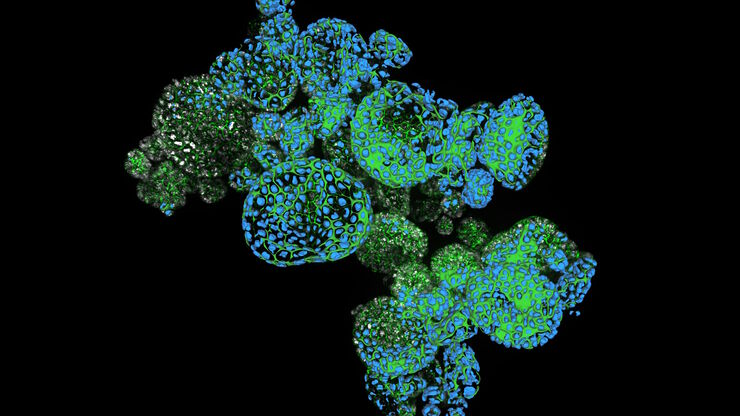
How Efficient is your 3D Organoid Imaging and Analysis Workflow?
Organoid models have transformed life science research but optimizing image analysis protocols remains a key challenge. This webinar explores a streamlined workflow for organoid research, starting…
Loading...

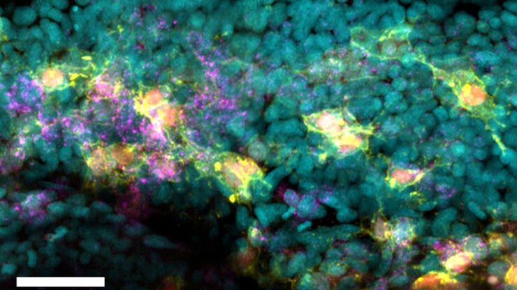
How do Cells Talk to Each Other During Neurodevelopment?
Professor Silvia Capello presents her group’s research on cellular crosstalk in neurodevelopmental disorders, using models such as cerebral organoids and assembloids.
Loading...

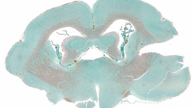
How to Image Histological and Fluorescent Samples with One System
VIDEO ON DEMAND - How to image histological and fluorescent samples with one system. FluoSync, the new technology embedded into Mica enables the imaging of both histological staining and fluorescence…
Loading...

How to Radically Simplify Workflows in Your Imaging Facility
VIDEO ON DEMAND - How to radically simplify imaging workflows and generate meaningful results with less time and effort using a highly automated microscope that unites widefield and confocal imaging.
Loading...

Advancing Surgery in Pediatric Brain Tumors
Learn how fluorescence-guided surgery supports pediatric brain tumor resection and improves precision through literature review and clinical cases.
Loading...

Alternative Fuels and Why Sustainable Solutions are Important
This free on-demand webinar is about the role of alternative fuel vehicles and why sustainable solutions are of increasing importance to the automotive industry.
