Webinars
Take a look at our upcoming and on-demand webinars. Join us at one of our next events!
Take a look at all our upcoming congresses, exhibitions, webinars, and workshops and join us at one of our next events!
22
–
23
Apr
2025
第四届半导体封装检测及失效分析技术进展网络研讨会
China
•
Webinar
22
Apr
2025
共聚焦荧光寿命成像功能在植物相关研究中的应用
China
•
Webinar
Filter articles
Tags
Products
Loading...
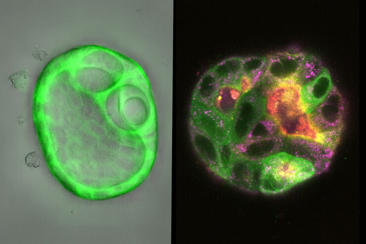
 and CellEvent™ (yellow).")
Following Multiple Events during Staurosporine Apoptosis
In this video on demand, we show how adding additional markers to an apoptosis kit can markedly increase the amount of information a researcher can obtain from the same experiment. The simultaneous…
Loading...
 stained to show the nucleus")
3D Spatial Analysis Using Mica's AI-Enabled Microscopy Software
This video offers practical advice on the extraction of publication grade insights from microscopy images. Our special guest Luciano Lucas (Leica Microsystems) will illustrate how Mica’s AI-enabled…
Loading...

Imaging of Cardiac Tissue Regeneration in Zebrafish
Learn how to image cardiac tissue regeneration in zebrafish focusing on cell proliferation and response during recovery with Laura Peces-Barba Castaño from the Max Planck Institute.
Loading...
How Does The Cytoskeleton Transport Molecules?
VIDEO ON DEMAND - See how 3D cysts derived from MDCK cells help scientists understand how proteins are transported and recycled in tissues and the role of the cytoskeleton in this transport.
Loading...
 embryo, from sphere stage to somite stages.")
Studying Early Phase Development of Zebrafish Embryos
This video on demand focuses on combining widefield and confocal imaging to study the early-stage development of zebrafish embryos (Danio rerio), from oocyte to multicellular stage.
Loading...

How To Get Multi Label Experiment Data With Full Spatiotemporal Correlation
This video on demand focuses on the special challenges of live cell experiments. Our hosts Lynne Turnbull and Oliver Schlicker use the example of studying the mitochondrial activity of live cells.…
