Webinars
Take a look at our upcoming and on-demand webinars. Join us at one of our next events!
Take a look at all our upcoming congresses, exhibitions, webinars, and workshops and join us at one of our next events!
22
–
23
Apr
2025
第四届半导体封装检测及失效分析技术进展网络研讨会
China
•
Webinar
22
Apr
2025
共聚焦荧光寿命成像功能在植物相关研究中的应用
China
•
Webinar
Filter articles
Tags
Products
Loading...
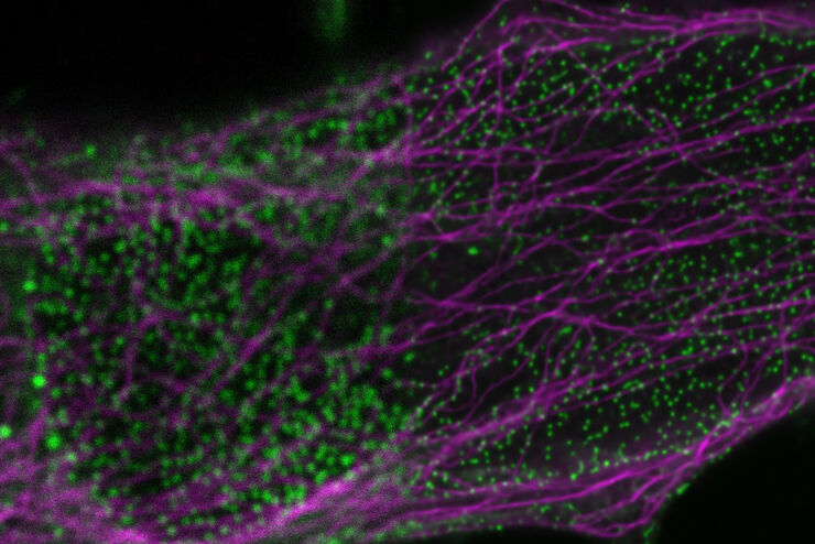
Tracking Single Cells Using Deep Learning
AI-based solutions continue to gain ground in the field of microscopy. From automated object classification to virtual staining, machine and deep learning technologies are powering scientific…
Loading...

Learning the Cellular Architecture from its Optical Properties
In the last 3 years, microscopists have started to use "AI based" solutions for a wide range of applications, including image acquisition optimization (smart microscopy), object classification, image…
Loading...
AI in Microscopy Webinar
We demonstrate residual channel attention networks for restoring and enhancing volumetric time-lapse (4D) fluorescence microscopy data.
Loading...

How to Conduct Standard-Compliant Analysis of Non-Metallic Inclusions in Steel
This webinar will provide an overview of the significance of non-metallic inclusions in steel and outline the important global standards for rating the quality of steel and difficulties that arise in…
Loading...

Industrial Microscopy: Digital imaging and the Leica DVM6
This webinar will discuss digital microscopy and Leica’s digital DVM6 microscope. We will navigate the difference between optical and digital magnification, explain the differences in optics, and…
Loading...
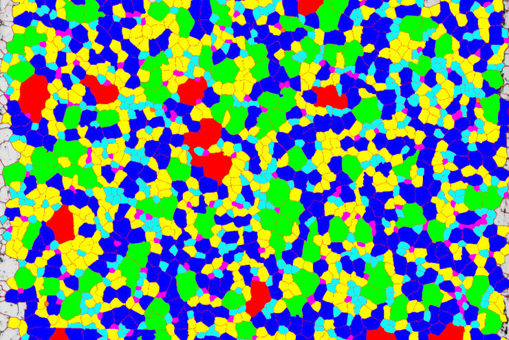
Inverted Microscopes for Grain Size Analysis: Three Factors to Consider
Microscopic steel grain size analysis is useful in determining the quality of steel alloys for a given purpose such as building bridges vs railroad rails. This webinar will describe the preparation of…
