Mateo
Inverse Mikroskope
Lichtmikroskope
Produkte
Startseite
Leica Microsystems
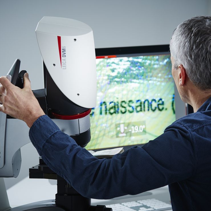
Mateo Digitalmikroskope
Entdecken Sie Mateo Familie
Lesen Sie unsere neuesten Artikel

Introduction to 21 CFR Part 11 for Electronic Records of Cell Culture
This article provides an introduction to the recommendations of 21 CFR Part 11 from the FDA, specifically focusing on the audit trail and user management in the context of cell-culture laboratories.…
.")
How Efficient is your 3D Organoid Imaging and Analysis Workflow?
Organoid models have transformed life science research but optimizing image analysis protocols remains a key challenge. This webinar explores a streamlined workflow for organoid research, starting…
 and analyzed for confluency using AI (right).")
AI Confluency Analysis for Enhanced Precision in 2D Cell Culture
This article explains how efficient, precise confluency assessment of 2D cell culture can be done with artificial intelligence (AI). Assessing confluency, the percentage of surface area covered,…

Precision and Efficiency with AI-Enhanced Cell Counting
This article describes the use of artificial intelligence (AI) for precise and efficient cell counting. Accurate cell counting is important for research with 2D cell cultures, e.g., cellular dynamics,…
 of U2OS cells which were transfected with a fluorescently labelled protein. A fluorescence image of the cells (right) is also shown. The analysis and imaging were performed with Mateo FL.")
Leveraging AI for Efficient Analysis of Cell Transfection
This article explores the pivotal role of artificial intelligence (AI) in optimizing transfection efficiency measurements within the context of 2D cell culture studies. Precise and reliable…

Overcoming Observational Challenges in Organoid 3D Cell Culture
Learn how to overcome challenges in observing organoid growth. Read this article and discover new solutions for real-time monitoring which do not disturb the 3D structure of the organoids over time.

An Introduction to Fluorescence
This article gives an introduction to fluorescence and photoluminescence, which includes phosphorescence, explains the basic theory behind them, and how fluorescence is used for microscopy.
 mit Phasenkontrast.")
Phasenkontrast und Mikroskopie
Dieser Artikel erklärt den Phasenkontrast, eine optische Mikroskopietechnik, die feine Details von ungefärbten, transparenten Proben sichtbar macht, die mit gewöhnlicher Hellfeldbeleuchtung schwer zu…

How to Determine Cell Confluency with a Digital Microscope
This article shows how to measure cell confluency in an easy and consistent way with Mateo TL, increasing confidence in downstream experiments.

How to do a Proper Cell Culture Quick Check
In order to successfully work with mammalian cell lines, they must be grown under controlled conditions and require their own specific growth medium. In addition, to guarantee consistency their growth…

Microscopy in Virology
The coronavirus SARS-CoV-2, causing the Covid-19 disease effects our world in all aspects. Research to find immunization and treatment methods, in other words to fight this virus, gained highest…

Introduction to Mammalian Cell Culture
Mammalian cell culture is one of the basic pillars of life sciences. Without the ability to grow cells in the lab, the fast progress in disciplines like cell biology, immunology, or cancer research…

Introduction to Widefield Microscopy
This article gives an introduction to widefield microscopy, one of the most basic and commonly used microscopy techniques. It also shows the basic differences between widefield and confocal…
Digitalmikroskope
Digitalmikroskope sind Mikroskope ohne Okulare. Eine Digitalkamera fungiert als Detektor. Bilder werden auf einem Monitor angezeigt – so wird der Mikroskopie-Arbeitsplatz zum ergonomischen…
Fields of Application
Zellkultur
Das Züchten von Zellkulturen unter Laborbedingungen ist die Basis wissenschaftlichen Arbeitens in der Zellbiologie, Krebsforschung, Entwicklungsbiologie oder jeder Art von biowissenschaftlicher und…
