is mobile? false
Science Lab
Science Lab
Willkommen auf dem Wissensportal von Leica Microsystems. Hier finden Sie wissenschaftliches Forschungs- und Lehrmaterial rund um das Thema Mikroskopie. Das Portal unterstützt Anfänger, erfahrene Praktiker und Wissenschaftler gleichermaßen bei ihrer täglichen Arbeit und ihren Experimenten. Erkunden Sie interaktive Tutorials und Anwendungshinweise, entdecken Sie die Grundlagen der Mikroskopie ebenso wie High-End-Technologien. Werden Sie Teil der Science Lab Community und teilen Sie Ihr Fachwissen.
Wissensportal Show subnavigation

Science Lab
Wissens- und Lernportal für Mikroskopie. Lernen. Teilen. Mitmachen.


Wählen Sie Ihr Anwendungsgebiet Show subnavigation


Filter articles
Tags
Berichtstyp
Produkte
Loading...
 and astrocytes (green) in a cortical spheroid derived from human induced pluripotent stem cells.")
Download The Guide to Live Cell Imaging
In life science research, live cell imaging is an indispensable tool to visualize cells in a state as in vivo as possible. This E-book reviews a wide range of important considerations to take to…
Loading...

Exploring the Structure and Life Cycle of Viruses
The SARS-CoV-2 outbreak started in late December 2019 and has since reached a global pandemic, leading to a worldwide battle against COVID-19. The ever-evolving electron microscopy methods offer a…
Loading...

Regulators of Actin Cytoskeletal Regulation and Cell Migration in Human NK Cells
Dr. Mace will describe new advances in our understanding of the regulation of human NK cell actin cytoskeletal remodeling in cell migration and immune synapse formation derived from confocal and…
Loading...

Adding Dimensions to Multiplex Molecular Imaging
Molecular imaging of living specimens offers a means to draw upon the growing body of high-throughput molecular data to better understand the underlying cellular and molecular mechanisms of complex…
Loading...
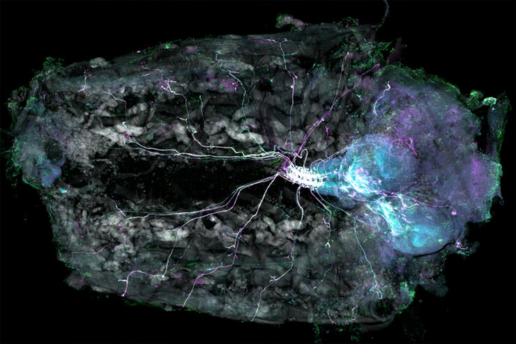
Understanding Motor Sequence Generation Across Spatiotemporal Scales
We have developed a microscopy-based pipeline to characterize a developmentally critical behavior at the pupal stage of development, called the ecdysis sequence. We study brain-wide neuronal activity…
Loading...

Benefits of TauContrast to Image Complex Samples
In this interview, Dr. Timo Zimmermann talks about his experience with the application of TauSense tools and their potential for the investigation of demanding samples such as thick samples or…
Loading...

Why is Manual Visual Inspection of Medical Devices so Challenging?
This article discusses how manual visual inspection, which is prevalent in the medical device industry, can lead to inconsistent results. It also addresses the challenges quality managers and…
Loading...
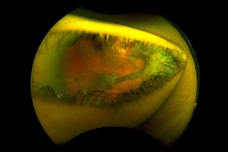
Intraoperative OCT-Assisted Surgical Management of Proliferative Vitreoretinopathy
Proliferative vitreoretinopathy (PVR) is a plague to patients and their surgeons after recent rhegmatogenous retinal detachment (RD). Despite excellent initial surgical outcomes, it is the most common…
Loading...

Fast, High-quality Vitrification with the EM ICE High Pressure Freezer
The EM ICE High Pressure Freezer was developed with a unique freezing principle and uses only a single pressurization and cooling liquid: liquified nitrogen (LN2). This design enables three major…
