Corporate Communications

Leica Microsystems entwickelt und fertigt Mikroskope und wissenschaftliche Instrumente für die Analyse von Mikro- und Nanostrukturen.
Wir bieten wissenschaftliches Forschungs- und Lehrmaterial zu den Themen der Mikroskopie an. Die Inhalte sind so gestaltet, dass sie Anfänger, erfahrene Praktiker und Wissenschaftler gleichermaßen bei ihrer täglichen Arbeit und ihren Experimenten unterstützen. Erkunden Sie interaktive Tutorials und Anwendungshinweise, entdecken Sie die Grundlagen der Mikroskopie ebenso wie High-End-Technologien.
Folgen Sie uns!

Unlocking the Secrets of Organoid Models in Biomedical Research
Get ready to delve deeper into the world of organoids and 3D models, which are essential tools for advancing our understanding of human health. Navigating these complex structures and obtaining clear…
 applied. Image courtesy of Samuel East, Uncommon Bio.")
Designing the Future with Novel and Scalable Stem Cell Culture
Visionary biotech start-up Uncommon Bio is tackling one of the world’s biggest health challenges: food sustainability. In this webinar, Stem Cell Scientist Samuel East shows how they make stem cell…

Mica: A Game-changer for Collaborative Research at Imperial College London
This interview highlights the transformative impact of Mica at Imperial College London. Scientists explain how Mica has been a game-changer, expanding research possibilities and facilitating…

Krebsforschung
Krebs ist eine komplexe und heterogene Krankheit, die durch Zellen verursacht wird, denen die Wachstumsregulation fehlt. Genetische und epigenetische Veränderungen in einer Zelle oder einer Gruppe von…
Phasenkontrast
Mit einem Phasenkontrast-Lichtmikroskop können die Strukturen vieler Arten von biologischen Präparaten mit einem größeren Kontrast betrachtet werden, ohne dass die Probe eingefärbt werden muss.
, and trafficking vesicles labeled with CF594 (cyan - Biotium).")
A Guide to Super-Resolution
Find out more about Leica super-resolution microscopy solutions and how they can empower you to visualize in fine detail subcellular structures and dynamics.

Präpariermikroskope
Wenn Sie Präparationen durchführen, verbringen Sie oft viele Stunden an den Okularen eines Präpariermikroskops. Bei Leica Microsystems können Sie aus einer Vielzahl von Mikroskopen und einem…

Neurowissenschaften
Arbeiten Sie an einem besseren Verständnis neurodegenerativer Erkrankungen oder an einer Untersuchung der Funktionen des Nervensystems? Erfahren Sie, wie Sie mit Bildgebungslösungen von Leica…

Virologie
Liegt Ihr Forschungsschwerpunkt auf Virusinfektionen und -krankheiten? Erfahren Sie, wie Sie mit Lösungen für Bildgebung und Probenvorbereitung von Leica Microsystems mehr Erkenntnisse in der…
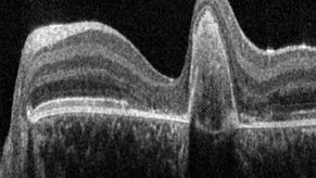
A Guide to OCT
Leica Optical Coherence Tomography (OCT) systems support ophthalmologists, ophthalmic surgeons, and researchers with easy-to-use, high-quality imaging technology.

Kryo-Elektronen-Tomographie
Mit der Kryo-Elektronentomographie (CryoET) lassen sich Biomoleküle in ihrer zellulären Umgebung mit einer noch nie dagewesenen Auflösung von weniger als einem Nanometer auflösen.

Zebrafisch-Forschung
Für optimale Ergebnisse während der Bewertung, Sortierung, Manipulation und Bildgebung von Modellorganismen ist es entscheidend feine Details und Strukturen genauestens zu erkennen. Das bildet die…

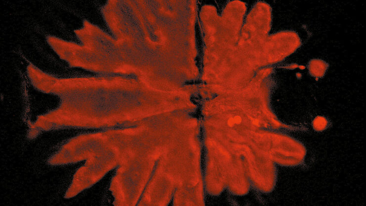
How to Study Gene Regulatory Networks in Embryonic Development
Join Dr. Andrea Boni by attending this on-demand webinar to explore how light-sheet microscopy revolutionizes developmental biology. This advanced imaging technique allows for high-speed, volumetric…

Cutting-Edge Imaging Techniques for GPCR Signaling
With this webinar on-demand enhance your pharmacological research with our webinar on GPCR signaling and explore cutting-edge imaging techniques that aim to understand how GPCR signaling translates…

Revealing Neuronal Migration’s Molecular Secrets
Different approaches can be used to investigate neuronal migration to their niche in the developing brain. In this webinar, experts from The University of Oxford present the microscopy tools and…

Advancing Uterine Regenerative Therapies with Endometrial Organoids
Prof. Kang's group investigates important factors that determine the uterine microenvironment in which embryo insertion and pregnancy are successfully maintained. They are working to develop new…

Lipidomics Analysis of Sparse Cells based on Laser Microdissection
Delve into cellular intricacies with high-coverage targeted lipidomics analysis of sparse cells. This advanced method, integrating Laser Microdissection (LMD) and Liquid Chromatography-Mass…
 set-up.")
Augmented Reality: Transformation von neurochirurgischen Verfahren
In diesem E-Book lernen Sie die großartigen Vorteile kennen, die Augmented Reality (AR) der Neurochirurgie bringt. Dieser umfassende Leitfaden enthält erklärende Videos, beantwortet brennende Fragen…

Laser Microdissection Protocols for Tissue and Cell Isolation - Download free eBook
In this Bio-protocol Selections, we present a collection of open-access, detailed methods papers using LCM to purify and isolate tissues and cells from plants, mouse embryos, cancer cells, neurons,…

Workflow Solutions for Sample Preparation Methods for Material Science
This brochure presents and explains appropriate workflow solutions for the most frequently required sample preparation methods for material science samples.
.")
Dual-View LightSheet Microscope for Large Multicellular Systems
Visualizing the dynamics of complex multicellular systems is a fundamental goal in biology. To address the challenges of live imaging over large spatiotemporal scales, Franziska Moos et. al. present…

Super-Resolution Microscopy Image Gallery
Due to the diffraction limit of light, traditional confocal microscopy cannot resolve structures below ~240 nm. Super-resolution microscopy techniques, such as STED, PALM or STORM or some…

Extended Live-cell Imaging at Nanoscale Resolution
Extended live-cell imaging with TauSTED Xtend. Combined spatial and lifetime information allow super-resolution microscopy at extremely low light dose.
, actin network (ATTO 647N), and nuclear pore basket (CF 680R).")
The Guide to STED Sample Preparation
This guide is intended to help users optimize sample preparation for stimulated emission depletion (STED) nanoscopy, specifically when using the STED microscope from Leica Microsystems. It gives an…
Accelerating Discovery for Multiplexed Imaging of Diverse Tissues
Explore IBEX: Open-source multiplexed imaging. Join the collaborative IBEX Imaging Community for optimized tissue processing, antibody selection, and human atlas construction.

Notable AI-based Solutions for Phenotypic Drug Screening
Learn about notable optical microscope solutions for phenotypic drug screening using 3D-cell culture, both planning and execution, from this free, on-demand webinar.

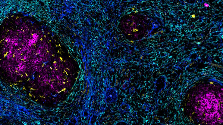
Understanding Tumor Heterogeneity with Protein Marker Imaging
Explore tumor heterogeneity and immune cell dynamics. See how quantitative imaging analysis reveals spatial relationships and molecular insights crucial for advancing cancer research and therapeutics.
 imaged with the THUNDER Imager 3D Cell Culture. Courtesy of Dr. F.T. Arroso Martins, Tamere University, Finland.")
How to Get Deeper Insights into your Organoid and Spheroid Models
In this eBook, learn about key considerations for imaging 3D cultures, such as organoids and spheroids, and discover microscopy solutions to shed new insights into dynamic processes in 3D real-time

Discover how Multiplexed Bioimaging can Advance Cancer Research
Explore multiplexing with up to 60 biomarkers, enabling advanced tumor imaging approaches to gather precise, spatially-resolved single-cell data that helps enhance cancer research and clinical…

Potential of Multiplex Confocal Imaging for Cancer Research and Immunology
Explore the new frontiers of multi-color fluorescent imaging: from image acquisition to analysis
