James DeRose , Ph.D.

James DeRose

James DeRose ist Global Marketing Communication Manager bei Leica Microsystems. Sein Schwerpunkt liegt auf der Erstellung optimierter Inhalte für Anwendungen in den Bereichen Life Science, Materialwissenschaften sowie Industrie und Fertigung. Er erstellt Applikationsberichte, Fallstudien, technische Berichte, White Papers, Anwendungsseiten, Produktseiten, Social Media Posts, E-Mails, Testimonials und andere Materialien. Er arbeitet seit 2013 bei Leica Microsystems. In der Vergangenheit arbeitete er an Anwendungsentwicklungsprojekten in den Bereichen Grenzflächenchemie und -physik, Wärme- und Verfahrenstechnik, Korrosion und Metallographie, Oberflächenbeschichtungen, Materialwissenschaften, Biotechnologie und Zellbiologie. Er hat Erfahrung mit verschiedenen Arten von Mikroskopie- und Analysemethoden.
Automated Laser Microdissection for Proteome Analysis

Deep Visual Proteomics Provides Precise Spatial Proteomic Information

Despite the availability of imaging methods and mass spectroscopy for spatial proteomics, a key challenge that remains is correlating images with single-cell resolution to protein-abundance…
These images show the microstructure of a hard metal with 10% cobalt which is used for heavy-duty tools. The large increase in magnification of the right image (compared to the left) has a risk of being outside the useful range or, in other words, empty magnification.

What is Empty Magnification and How can Users Avoid it

The phenomenon of “empty magnification”, which can occur while using an optical, light, or digital microscope, and how it can be avoided is explained in this article. The performance of an optical…
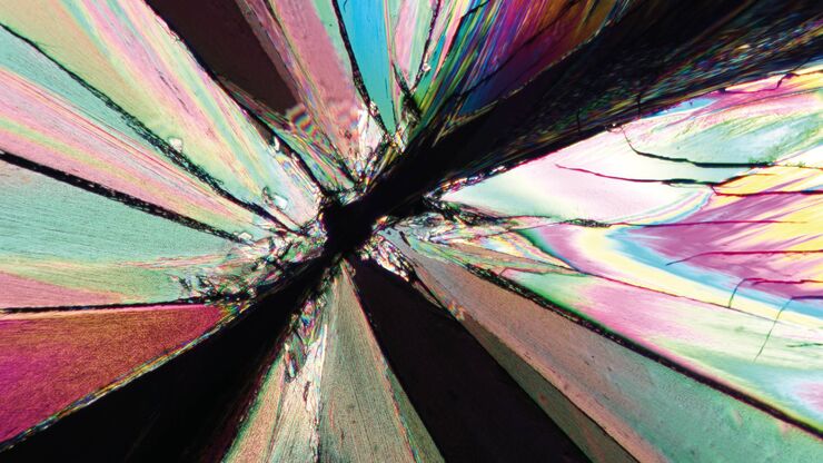
Image of a tartaric-acid crystal taken with polarization microscopy. Tartaric acid, a diprotic, aldaric carboxylic acid, is a naturally occurring organic compound notably found in grapes.

Das Prinzip der Polarisationsmikroskopie

Die Polarisationsmikroskopie wird in den Material- und Geowissenschaften routinemäßig eingesetzt, um Materialien und Mineralien anhand ihrer charakteristischen Brechungseigenschaften und Farben zu…
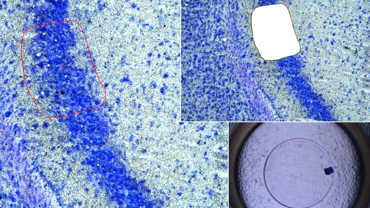
Mouse brain (left) microdissected with a 10x objective (upper right). Inspection of the collection device (lower right).

Molecular Biology Analysis facilitated with Laser Microdissection (LMD)

Extracting biomolecules, proteins, nucleic acids, lipids, and chromosomes, as well as extracting and manipulating cells and tissues with laser microdissection (LMD) enables insights to be gained into…
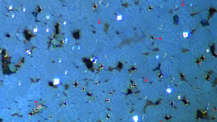
Image of magnetic steel taken with a 100x objective using Kerr microscopy. The magnetic domains in the grains appear in the image with lighter and darker patterns. A few domains are marked with red arrows. Courtesy of Florian Lang-Melzian, Robert Bosch GmbH, Germany.

Rapidly Visualizing Magnetic Domains in Steel with Kerr Microscopy

The rotation of polarized light after interaction with magnetic domains in a material, known as the Kerr effect, enables the investigation of magnetized samples with Kerr microscopy. It allows rapid…
AI-based transfection analysis (left) of U2OS cells which were transfected with a fluorescently labelled protein. A fluorescence image of the cells (right) is also shown. The analysis and imaging were performed with Mateo FL.

Leveraging AI for Efficient Analysis of Cell Transfection

This article explores the pivotal role of artificial intelligence (AI) in optimizing transfection efficiency measurements within the context of 2D cell culture studies. Precise and reliable…
AI-based cell counting performed with a phase-contrast and fluorescence image using the Mateo FL microscope.

Precision and Efficiency with AI-Enhanced Cell Counting

This article describes the use of artificial intelligence (AI) for precise and efficient cell counting. Accurate cell counting is important for research with 2D cell cultures, e.g., cellular dynamics,…
Image of confluent cells taken with phase contrast (left) and analyzed for confluency using AI (right).

AI Confluency Analysis for Enhanced Precision in 2D Cell Culture

This article explains how efficient, precise confluency assessment of 2D cell culture can be done with artificial intelligence (AI). Assessing confluency, the percentage of surface area covered,…
Image of murine dopaminergic neurons which have been marked for laser microdissection (LMD).

Neuron Isolation in Spatial Context with Laser Microdissection (LMD)

After Alzheimer’s disease, Parkinson’s is the second most common progressive neurodegenerative disease. Before the first symptoms manifest, up to 70% of dopamine-releasing neurons in the mid-brain…
Region of a patterned wafer inspected using optical microscopy and automated and reproducible DIC (differential interference contrast). With DIC users are able to visualize small height differences on the wafer surface more easily.

6-Inch Wafer Inspection Microscope for Reliably Observing Small Height Differences

A 6-inch wafer inspection microscope with automated and reproducible DIC (differential interference contrast) imaging, no matter the skill level of users, is described in this article. Manufacturing…
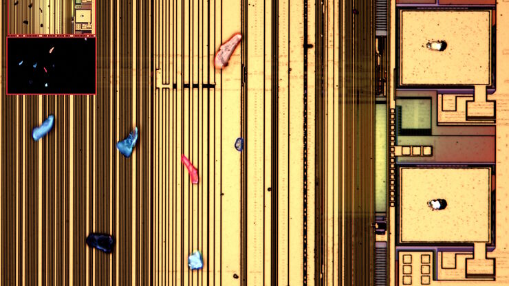
Optical microscope image, which is a composition of both brightfield and fluorescence illumination, showing organic contamination on a wafer surface. The inset images in the upper left corner show the brightfield image (above) and fluorescence image (below with dark background).

Visualizing Photoresist Residue and Organic Contamination on Wafers

As the scale of integrated circuits (ICs) on semiconductors passes below 10 nm, efficient detection of organic contamination, like photoresist residue, and defects during wafer inspection is becoming…
Eine Batterieelektrode, deren Ränder Grate aufweisen (mit roten Pfeilen markiert). Das Bild wurde mit einem Digitalmikroskop DVM6 aufgenommen.

Graterkennung während der Batterieherstellung

Erfahren Sie, wie die optische Mikroskopie zur Graterkennung an Batterieelektroden und zur Bestimmung des Schadenspotenzials eingesetzt werden kann, um eine schnelle und zuverlässige…
Partikel auf der Oberfläche einer Partikelfalle, die für die technische Sauberkeit bei der Batterieproduktion eingesetzt werden kann.

Erkennung von Batteriepartikeln während des Produktionsprozesses

In diesem Artikel wird erläutert, wie die Partikelerkennung und -analyse von Batterien mit optischer Mikroskopie und Laserspektroskopie für eine schnelle, zuverlässige und kostengünstige…
THUNDER image of brain-capillary endothelial-like cells derived from human iPSCs (induced pluripotent stem cells) where cyan indicates nuclei and magenta tight junctions.

Rapid Check of Live Stem Cells in Cell-Culture Inserts set in Multi-Well Plates

See how efficient imaging of live iPSC stem cells within cell-culture inserts set in a multi-well plate can be done to evaluate the cells using a THUNDER Imager. Just read this article.
Particulate contamination in between moving metal plates.

Key Factors for Efficient Cleanliness Analysis

An overview of the key factors necessary for technical cleanliness and efficient cleanliness analysis concerning automotive and electronics manufacturing and production is provided in this article.
Microscope equipped with a K7 color CMOS camera for life-science and industry imaging applications.

Technical Terms for Digital Microscope Cameras and Image Analysis

Learn more about the basic principles behind digital microscope camera technologies, how digital cameras work, and take advantage of a reference list of technical terms from this article.
Images of the same area of a processed wafer taken with standard (left) and oblique (right) brightfield illumination using a Leica compound microscope. The defect on the wafer surface is clearly more visible with oblique illumination.

Rapid Semiconductor Inspection with Microscope Contrast Methods

Semiconductor inspection during the production of patterned wafers and ICs (integrated circuits) is important for identifying and minimizing defects. To increase the efficiency of quality control in…
Preparation of an IC-chip cross section: grinding and polishing of the chip cross section.

Cross-section Analysis for Electronics Manufacturing

This article describes cross-section analysis for electronics concerning quality control and failure analysis of printed circuit boards (PCBs) and assemblies (PCBAs), integrated circuits (ICs), etc.
Bild von immunfluoreszierend markierten Zellen, wobei Mitochondrien rot, Zellkerne blau und Aktin grün dargestellt sind.

Erforschung der Virusreplikaton mit Fluoreszenzmikroskopie

Viren können mit Hilfe verschiedener Mikroskopietechniken untersucht werden. Je nach Vergrößerung und Auflösung des Mikroskops kann die Beobachtung auf Gewebe-, Zell- oder Virionenebene erfolgen.
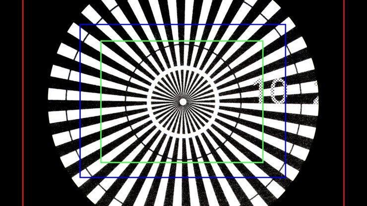
Image of a Siemens star, where the diameter of the 1st black line circle is 10 mm and the 2nd is 20 mm, taken via an eyepiece of a M205 A stereo microscope. The rectangles represent the field of view (FOV) of a Leica digital camera when installed with various C-mounts (red 0.32x, blue 0.5x, green 0.63x).

Understanding Clearly the Magnification of Microscopy

To help users better understand the magnification of microscopy and how to determine the useful range of magnification values for digital microscopes, this article provides helpful guidelines.
Fluorescence microscopy image of liver tissue where DNA in the nuclei are stained with Feulgen-pararosanilin and visualized with transmitted green light.

Epi-Illumination Fluorescence and Reflection-Contrast Microscopy

This article discusses the development of epi-illumination and reflection contrast for fluorescence microscopy concerning life-science applications. Much was done by the Ploem research group…
Image of an onion flake taken with a basic Leica compound microscope after it was tested for resistance to fungus and mold growth following part 11 of the ISO 9022 standard.

ISO 9022 Standard Part 11 - Testing Microscopes with Severe Conditions

This article describes a test to determine the robustness of Leica microscopes to mold and fungus growth. The test follows the specifications of the ISO 9022 part 11 standard for optical instruments.
EBSD-Korngrößenverteilung des Querschnitts eines Golddrahtes in einer Siliziummatrix aus dem Inneren einer CPU (Zentraleinheit eines Computers). Die Körner sind mit verschiedenen Farben hervorgehoben.

Hochwertige EBSD-Probenvorbereitung

Es wird eine zuverlässige und effiziente EBSD-Probenvorbereitung von „gemischten“ kristallografischen Materialien mit Ionenbreitstrahlfräsen beschrieben. Das beschriebene Verfahren erzeugt…
Stereomikroskope werden oft als die Arbeitstiere von Labors und Produktionsstätten angesehen.

Wichtige Faktoren, die Sie bei der Auswahl eines Stereomikroskops berücksichtigen sollten

Stereomikroskope zeichnen sich durch ihre Fähigkeit aus, einen 3D-Eindruck der Probe zu erzeugen. Daher eignen sie sich besonders gut für Inspektion und Nacharbeit, Qualitätskontrolle, Forschung und…
Geringe Vergrößerung DVM6-Bild eines Teils der PCBA-Probe.

Schnelle und zuverlässige Untersuchung von Leiterplatten und Leiterplattenbaugruppen mittels Digitalmikroskopie

Digitalmikroskope bieten Anwendern eine bequeme und schnelle Möglichkeit zur Erfassung hochwertiger, zuverlässiger Bilddaten und zur schnellen Inspektion und Analyse von Leiterplatten (PCBs) und…
Fluorescence microscope image of a life-science specimen

An Introduction to Fluorescence

This article gives an introduction to fluorescence and photoluminescence, which includes phosphorescence, explains the basic theory behind them, and how fluorescence is used for microscopy.
Raw widefield and THUNDER image of calcium transients in Drosophila embryos. Courtesy A. Carreira-Rosario, Clandinin laboratory, California, USA.

Central Nervous System (CNS) Development and Activity in Organisms

This article shows how studying central nervous system (CNS) development in Drosophila-melanogaster embryos expressing a GCaMP calcium indicator in the neurons can be improved with a THUNDER Imager.
Patch pipette touching a murine hippocampal neuron. Image courtesy of A. Aguado, Ruhr University Bochum, Germany.

What is the Patch-Clamp Technique?

This article gives an introduction to the patch-clamp technique and how it is used to study the physiology of ion channels for neuroscience and other life-science fields.
Neurons imaged with DIC contrast.

Differential Interference Contrast (DIC) Microscopy

This article demonstrates how differential interference contrast (DIC) can be actually better than brightfield illumination when using microscopy to image unstained biological specimens.
The Emspira 3 digital microscope offers what users need for comprehensive visual inspection, including comparison, measurement, and documentation sharing.

Digital Inspection Microscope for Industrial Applications

Factors users should consider before choosing a digital inspection microscope for industrial applications, including quality control (QC), failure analysis (FA), and R&D, are described in this…
Scroll to top