James DeRose , Ph.D.

James DeRose

James DeRose ist Global Marketing Communication Manager bei Leica Microsystems. Sein Schwerpunkt liegt auf der Erstellung optimierter Inhalte für Anwendungen in den Bereichen Life Science, Materialwissenschaften sowie Industrie und Fertigung. Er erstellt Applikationsberichte, Fallstudien, technische Berichte, White Papers, Anwendungsseiten, Produktseiten, Social Media Posts, E-Mails, Testimonials und andere Materialien. Er arbeitet seit 2013 bei Leica Microsystems. In der Vergangenheit arbeitete er an Anwendungsentwicklungsprojekten in den Bereichen Grenzflächenchemie und -physik, Wärme- und Verfahrenstechnik, Korrosion und Metallographie, Oberflächenbeschichtungen, Materialwissenschaften, Biotechnologie und Zellbiologie. Er hat Erfahrung mit verschiedenen Arten von Mikroskopie- und Analysemethoden.

Keeping Particulate Contamination Under Control in Pharmaceutical Products

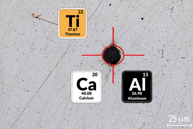
This article describes how a 2-methods-in-1 solution combining optical microscopy and laser induced breakdown spectroscopy (LIBS) can be utilized for identification of particulate contaminants in the…

Fast Visual and Chemical Analysis of Contamination and Underlying Layers

Visual and chemical analysis of contamination on materials with a 2-methods-in-1 solution leading to an efficient, more complete analysis workflow is described in this report. A 2-in-1 solution,…

How does an Automated Rating Solution for Steel Inclusions Work?

The rating of non-metallic inclusions (NMIs) to determine steel quality is critical for many industrial applications. For an efficient and cost-effective steel quality evaluation, an automated NMI…

Perform Microscopy Analysis for Pathology Ergonomically and Efficiently

The main performance features of a microscope which are critical for rapid, ergonomic, and precise microscopic analysis of pathology specimens are described in this article. Microscopic analysis of…

Challenges Faced When Manually Rating Non-Metallic Inclusions (NMIs) to Determine Steel Quality

Rapid, accurate, and reliable rating of non-metallic inclusions (NMIs) is instrumental for the determination of steel quality. This article describes the challenges that arise from manual NMI rating,…
Influenca in lung epithelial cells (porcine) - THUNDER Imager 3D Cell Culture Influenca virus – red, cilia – green, Nuclei – blue.

How Can Immunofluorescence Aid Virology Research?

Modern virology research has become as crucial now as ever before due to the global COVID-19 pandemic. There are many powerful technologies and assays that virologists can apply to their research into…

Reasons Why There is Growing Need for Fast and Reliable Steel Quality Rating Solutions

Steel quality is critical for the manufacturing of high-quality components and products. Fast, reliable, and accurate detection and classification of inclusions has become essential for both component…

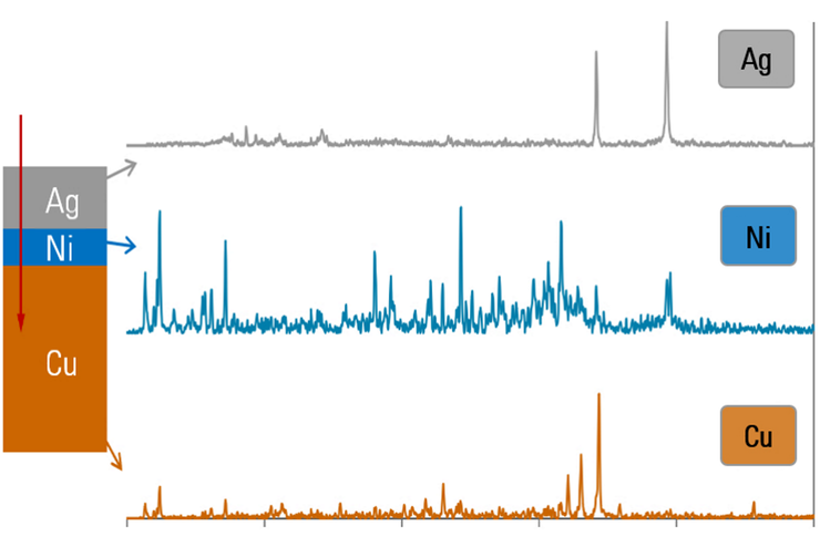
Depth Profiling and Layer Analysis for Inspection of Materials with a 2-In-1 Solution Combining Optical Microscopy and Laser Spectroscopy

In addition to simultaneous visual and chemical inspection, a 2-methods-in-1 materials analysis solution, which combines optical microscopy and laser induced breakdown spectroscopy (LIBS), can also be…

Top Issues Related to Standards for Rating Non-Metallic Inclusions in Steel

Supplying components and products made of steel to users worldwide can require that a single batch be compliant with multiple steel quality standards. This user demand creates significant challenges…

Visual and Chemical Analysis of Steel Microstructure: Faster Rating of Steel Quality

Simultaneous visual and chemical analysis of steel non-metallic inclusions with a 2-methods-in-1 solution, using optical microscopy and laser induced breakdown spectroscopy (LIBS), is described in…

Rate the Quality of Your Steel: Free Webinar and Report

This webinar and report describe optimal microscopy solutions for rating steel quality in terms of non-metallic inclusions and reviews the various international and regional standards concerning…

Metallography – an Introduction

This article gives an overview of metallography and metallic alloy characterization. Different microscopy techniques are used to study the alloy microstructure, i.e., microscale structure of grains,…

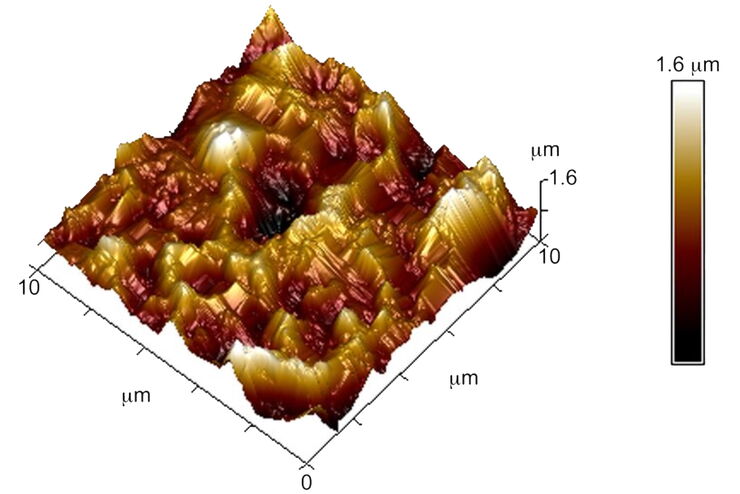
Brief Introduction to Surface Metrology

This report briefly discusses several important metrology techniques and standard definitions commonly used to assess the topography of surfaces, also known as surface texture or surface finish. With…

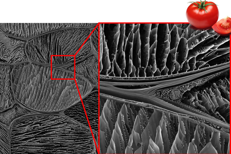
Studying the Microstructure of Natural Polymers in Fine Detail

The potential of cryogenic broad ion beam milling used in combination with scanning electron microscopy (cryo-BIB-SEM) for imaging and analyzing the microstructure of cryogenically stabilized soft…
Ultramicrotomy; ARTOS 3D

Introduction to Ultramicrotomy

When studying samples, to visualize their fine structure with nanometer scale resolution, most often electron microscopy is used. There are 2 types: scanning electron microscopy (SEM) which images the…
Measuring grains size with Abrams Three-Circle Procedure.

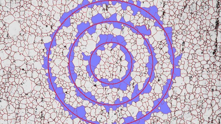
How to Adapt Grain Size Analysis of Metallic Alloys to Your Needs

Metallic alloys, such as steel and aluminum, have an important role in a variety of industries, including automotive and transportation. In this report, the importance of grain size analysis for alloy…

How to make a fast Z-stack

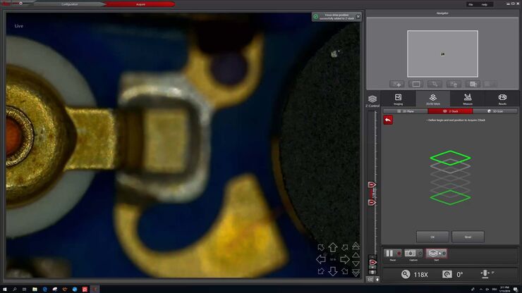
Save time for your 2D and 3D analysis. Watch this video to learn about the new user interface, LAS X.next, for the DVM6 digital microscope. The video demonstrates how to make a fast Z-Stack with a few…
Array tomography image of T-cells in mouse lymph nodes.

High Resolution Array Tomography with Automated Serial Sectioning

The optimization of high resolution, 3-dimensional (3D), sub-cellular structure analysis with array tomography using an automated serial sectioning solution, achieving a high section density on the…

Macroscale to Nanoscale Pore Analysis of Shale and Carbonate Rocks

Physical porosity in rocks, like shale and carbonate, has a large effect on the their storage capacity. The pore geometries also affect their permeability. Imaging the visible pore space provides…

See the Structure with Microscopy - Know the Composition with Laser Spectroscopy

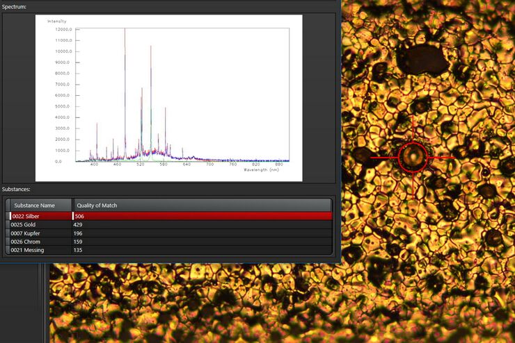
The advantages of a 2-in-1 materials analysis solution combining optical microscopy and laser induced breakdown spectroscopy (LIBS) for simultaneous visual and chemical inspection are described in…
Convalaria

Introduction to Widefield Microscopy

This article gives an introduction to widefield microscopy, one of the most basic and commonly used microscopy techniques. It also shows the basic differences between widefield and confocal…
Comparison when observing a rodent model organism with a Greenough versus CMO (common main objective) stereo microscope for a task like surgery.

Rodent and Small-Animal Surgery

Learn how you can perform rodent (mouse, rat, hamster) and small-animal surgery efficiently with a microscope for developmental biology and medical research applications by reading this article.
Transgenic zebrafish larva where fluorescent proteins label the heart muscle blue, blood and blood vessels red, and all circulatory system cells green. Image recorded with a M205 FA microscope.

Imaging and Analyzing Zebrafish, Medaka, and Xenopus

Discover how to image and analyze zebrafish, medaka, and Xenopus frog model organisms efficiently with a microscope for developmental biology applications from this article.
Fluorescence stereo microscope image of anesthetized Mediterranean fruit flies recorded with a M205 stereo microscope.

Investigating Fruit Flies (Drosophila melanogaster)

Learn how to image and investigate Drosophila fruit fly model organisms efficiently with a microscope for developmental biology applications from this article.
C. elegans

Studying Caenorhabditis elegans (C. elegans)

Find out how you can image and study C. elegans roundworm model organisms efficiently with a microscope for developmental biology applications from this article.

Life Science Imaging with DVM6 Digital Microscope

Digital microscopes can be a great help in life science applications such as the documentation in botany, entomology studies and crop science, or the digitization of museum collections. The image…
Watch imaged with DMS300.

What You Always Wanted to Know About Digital Microscopy, but Never Got Around to Asking

Digital microscopy is one of the buzz words in microscopy – and there are a couple of facts that are useful to know. Georg Schlaffer, Product Manager with Leica Microsystems, has often been asked…
Live cell imaging, 4 colors: Mitochondria (MitoView Green, yellow) and actin (mNeonGreen, cyan) microtubuli (SIR-tubulin, magenta), endosomes (NIR750, green). Processed with DSE and DSE powered by Aivia.

The Principles of White Light Laser Confocal Microscopy

The perfect light source for confocal microscopes in biomedical applications has sufficient intensity, tunable color and is pulsed for use in lifetime fluorescence. Furthermore, it should offer means…
An ID card which has been tampered with by counterfeiters who inserted a hologram.

Is that Document Genuine or Fake? How do They Identify Fake Documents?

This article shows how forensic experts use microscopy for analysis to identify counterfeit, fake documents, such as ID cards, passports, visas, certificates, etc. Then they know if it is genuine or…
A portion of an early binocular microscope developed by John Leonhard Riddel in the early 1850s.

The History of Stereo Microscopy

This article gives an overview on the history of stereo microscopes. The development and evolution from handcrafted instruments (late 16th to mid-18th century) to mass produced ones the last 150…
Scroll to top