Levi Felts , Dr.

Marketing Manager Microscopie, Leica Microsystems Inc., Buffalo Grove

Mouse kidney section with Alexa Fluor™ 488 WGA, Alexa Fluor™ 568 Phalloidin, and DAPI. Sample is a FluoCells™ prepared slide #3 from Thermo Fisher Scientific, Waltham, MA, USA. Images courtesy of Dr. Reyna Martinez – De Luna, Upstate Medical University, Department of Ophthalmology.

The Power of Pairing Adaptive Deconvolution with Computational Clearing

Learn how deconvolution allows you to overcome losses in image resolution and contrast in widefield fluorescence microscopy due to the wave nature of light and the diffraction of light by optical…
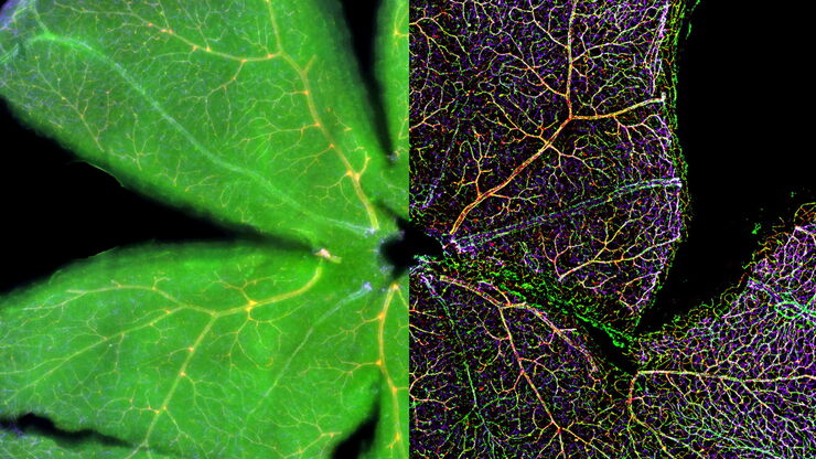
Mouse retina was fixed and stained by following reagents: anti-CD31 antibody (green): Endothelia cells, IsoB4 (red): Blood vessels, and microglia anti-GFAP antibody (blue): Astrocytes Sample courtesy by Jeremy Burton, PhD and Jiyeon Lee, PhD, Genentech Inc., South San Francisco, USA. Imaged by Olga Davydenko, PhD (Leica). Imaged with a THUNDER Imager 3D Cell Culture.

An Introduction to Computational Clearing

Many software packages include background subtraction algorithms to enhance the contrast of features in the image by reducing background noise. The most common methods used to remove background noise…
Scroll to top