Kontaktieren Sie uns

Lynne Turnbull

Lynne Turnbull

Lynne ist eine leitende Wissenschaftlerin bei Leica Microsystems. Sie promovierte in Sydney (Australien) im Bereich der Herzbiophysik und absolvierte eine Postdoc-Ausbildung in San Francisco und Melbourne. Lynnes Forschungsinteressen verlagerten sich auf bakterielle Biofilme und Motilität, und sie nutzte verschiedene Arten der Bildgebung, um zu erforschen und zu verstehen, wie Bakterien Gemeinschaften bilden und sich in ihrer Umgebung bewegen. Nach ihrem Wechsel an die University of Technology Sydney gründete und leitete Lynne die Microbial Imaging Facility. Lynne kam 2016 zu GE, um in ganz Asien Anwendungsunterstützung für hochauflösende Mikroskopie zu leisten.  Seit 2021 arbeitet Lynne bei Leica Microsystems in den Labors des EMBL Imaging Center in Heidelberg.

Multiplexed Cell DIVE imaging to characterize the spatial landscape in Human Alzheimer’s Cortical Tissue

Probing Human Alzheimer's Cortical Section using Spatial Multiplexing

Alzheimer’s disease (AD) is the most common neurodegenerative disease and is characterized by the progressive decline of cognitive function. Spatial profiling of AD brain may reveal cellular…
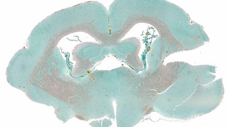
Masson-Goldner staining of a hedgehog brain slice.

How to Image Histological and Fluorescent Samples with One System

VIDEO ON DEMAND - How to image histological and fluorescent samples with one system. FluoSync, the new technology embedded into Mica enables the imaging of both histological staining and fluorescence…

How to Radically Simplify Workflows in Your Imaging Facility

VIDEO ON DEMAND - How to radically simplify imaging workflows and generate meaningful results with less time and effort using a highly automated microscope that unites widefield and confocal imaging.

Harnessing Microfluidics to Maintain Cell Health During Live-Cell Imaging

VIDEO ON DEMAND - In this webinar on-demand, we will use microfluidics to explore the effect of shear stress on cell morphology, examine the effect of nutrient replenishment on cellular growth during…
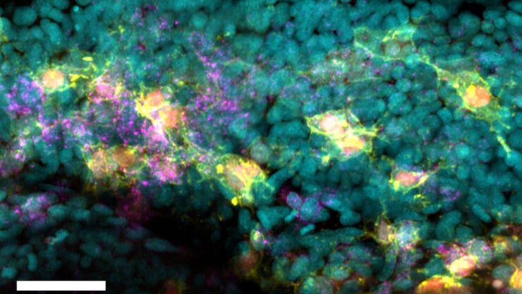
Two-color caspase assay with tile scan. U2OS cells were treated with the nuclear marker DRAQ5 (magenta) and CellEvent™ (yellow).

Following Multiple Events during Staurosporine Apoptosis

In this video on demand, we show how adding additional markers to an apoptosis kit can markedly increase the amount of information a researcher can obtain from the same experiment. The simultaneous…
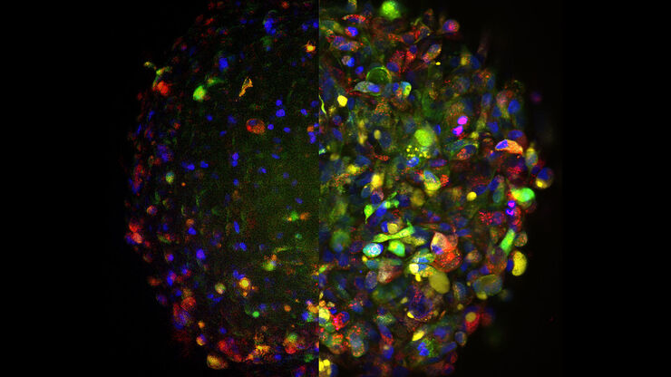
Untreated Hela Kyoto cells stained to show the nucleus (Hoechst, blue), the cis-golgi matrix protein GM130 (AF488, green), and the trans-golgi network membrane protein TGN46 (AF647, red).

Golgi Organizational Changes in Response to Cell Stress

In this video on demand, our special guest George Galea from EMBL Heidelberg will look at HeLa Kyoto cells treated with various chemotherapeutic agents to investigate their effect on the Golgi complex…
Protist Paramecium (Paramecium tetraurelia) stained to show the nucleus

AI-Enabled Spatial Analysis of Complex 3D Datasets

This video on demand offers practical advice on the extraction of publication grade insights from microscopy images. Our special guest Luciano Lucas (Leica Microsystems) will illustrate how Mica’s…
Image of a single slice taken from a zebrafish heart showing the ventricle with an injury in the lower area. Nuclei of all cells are indicated with blue, nuclei of the cardiomyocyte heart muscle cells with green, and the proliferating cells with red. Courtesy of Laura Peces-Barba Castaño, Max Planck Institute for Heart and Lung Research, Germany.

Imaging of Cardiac Tissue Regeneration in Zebrafish

Learn how to image cardiac tissue regeneration in zebrafish focusing on cell proliferation and response during recovery with Laura Peces-Barba Castaño from the Max Planck Institute.
U2OS cells stained with Hoechst for nuclei (blue), MitoTracker green (Mitochondria structure, green) and TMRE (active mitochondria, magenta) and SiR for tubulin (red). Simultaneous acquisition of four channel large area overview using Spiral scan feature using the 10x/1.20 CS2 Water MotCORR objective.

How To Get Multi Label Experiment Data With Full Spatiotemporal Correlation

This video on demand focuses on the special challenges of live cell experiments. Our hosts Lynne Turnbull and Oliver Schlicker use the example of studying the mitochondrial activity of live cells.…
Scroll to top