Peter Laskey , Dr.

Peter Laskey

Pete Laskey studierte Biologische und Medizinische Chemie, absolvierte dann einen Master in Pharmakologie und promovierte in Virologie. Während seiner Promotion untersuchte er die Wechselwirkung zwischen dem E1^E4-Protein des humanen Papillomavirus und dem Zytoskelett der Zellen und spezialisierte sich auf die Bildgebung von lebenden Zellen. Anschließend arbeitete er als Bildgebungsspezialist am National Institute of Medical Research, bevor er für mehrere Jahre die Welt bereiste. Nach seiner Rückkehr nach Europa verbrachte er mehrere Jahre bei Andor, wo er sich auf die sCMOS-Kameratechnologie konzentrierte, bevor er 2013 als Produktmanager für das GSD 3D Super Resolution System zu Leica wechselte.

Microscope equipped with a K7 color CMOS camera for life-science and industry imaging applications.

Technical Terms for Digital Microscope Cameras and Image Analysis

Learn more about the basic principles behind digital microscope camera technologies, how digital cameras work, and take advantage of a reference list of technical terms from this article.
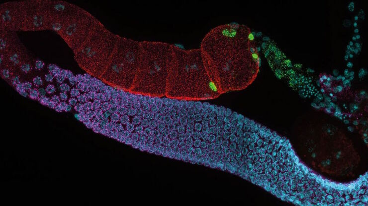
Ausgewachsene hermaphroditische Gonaden von C. elegans, aufgenommen mit THUNDER Imager. Färbung: blau - DAPI (Zellkern), grün - SP56 (Spermien), rot - RME-2 (Eizelle), magenta - PGL-1 (RNA + Proteingranula). Bild mit freundlicher Genehmigung von Prof. Dr. Christian Eckmann, Martin-Luther-Universität, Halle, Deutschland.

Life-Science-Forschung: Welche Mikroskopkamera ist die richtige für Sie?

Wie Sie entscheiden, welche Kamera für Ihre Life-Science-Mikroskopie-Experimente die Richtige ist. Welche Kamera von Leica Microsystems ist für Sie am besten geeignet?
Staining is crucial to revealing details in the specimens.

H&E Staining in Microscopy

If we consider the role of microscopy in pathologists’ daily routines, we often think of the diagnosis. While microscopes indeed play a crucial role at this stage of the pathology lab workflow, they…
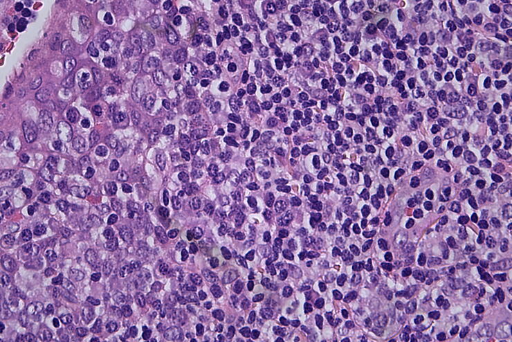
Histopathological sample, 40x magnification

Clinical Microscopy: Considerations on Camera Selection

The need for images in pathology laboratories has significantly increased over the past few years, be it in histopathology, cytology, hematology, clinical microbiology, or other applications. They…
Scroll to top