Volker Schweikhard , Dr.

Volker Schweikhard

Dr. Volker Schweikhard promovierte in Physik an der University of Colorado in Boulder und entwickelte optische Manipulationstechniken für ultrakalte Quantengase. Danach verlagerte er seinen Schwerpunkt auf die Forschung in den Biowissenschaften und untersuchte als Postdoc an der Stanford University den Prozess der Gentranskription auf Einzelmolekülebene. Als Research Assistant Professor in der Abteilung für Bioengineering an der Rice University in Houston, TX, erforschte er die subzelluläre Koordination von Zytoskelett-Netzwerken und Signalkaskaden.

Mit seinem Hintergrund in Optik, Biophysik und Zellbiologie ist es seine Leidenschaft, Instrumente und Verfahren zur Untersuchung biologischer Prozesse auf (sub-)zellulärer und Gewebeebene zu entwickeln. Bei Leica Microsystems arbeitet er mit Kunden, Kooperationspartnern und der internen Forschung und Entwicklung zusammen, um Innovationen in der nichtlinearen optischen Mikroskopie und der multimodalen Bildgebung voranzutreiben, einschließlich der Coherent anti-Stokes Raman Scattering Mikroskopie (CARS, SRS), der Multiphotonen-Fluoreszenzmikroskopie und der zeitaufgelösten Bildgebung.

An 8-color spectral unmixing result from a hyperspectral SRS (stimulated Raman scattering) dataset, showing the biochemically distinct structures of a fresh, untreated apple slice.

How to Prepare Samples for Stimulated Raman Scattering (SRS) imaging

Find here guidelines for how to prepare samples for stimulated Raman scattering (SRS), acquire images, analyze data, and develop suitable workflows. SRS spectroscopic imaging is also known as SRS…

Coherent Raman Scattering Microscopy Publication List

CRS (Coherent Raman Scattering) microscopy is an umbrella term for label-free methods that image biological structures by exploiting the characteristic, intrinsic vibrational contrast of their…
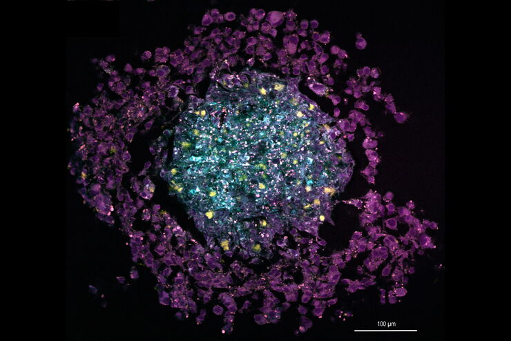
Multi-color SRS image of a tri-cellular cancer spheroid

The Potential of Coherent Raman Scattering Microscopy at a Glance

Coherent Raman scattering microscopy (CRS) is a powerful approach for label-free, chemically specific imaging. It is based on the characteristic intrinsic vibrational contrast of molecules in the…

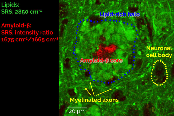
Stimulated Raman Scattering Microscopy Probes Neurodegenerative Disease

Despite decades of research, the molecular mechanisms underlying some of the most severe neurodegenerative diseases, such as Alzheimer’s or Parkinson’s, remain poorly understood. The progression of…
Scroll to top