
is mobile? false
Biowissenschaften
Biowissenschaften
Hier können Sie Ihr Wissen, Ihre Forschungsfähigkeiten und Ihre praktischen Anwendungen der Mikroskopie in verschiedenen wissenschaftlichen Bereichen erweitern. Erfahren Sie, wie Sie präzise Visualisierung, Bildinterpretation und Forschungsfortschritte erzielen können. Hier finden Sie aufschlussreiche Informationen über fortgeschrittene Mikroskopie, Bildgebungsverfahren, Probenvorbereitung und Bildanalyse. Zu den behandelten Themen gehören Zellbiologie, Neurowissenschaften und Krebsforschung mit Schwerpunkt auf modernsten Anwendungen und Innovationen.
Beliebt Show subnavigation
Biowissenschaften


Filter articles
Tags
Berichtstyp
Produkte
Loading...

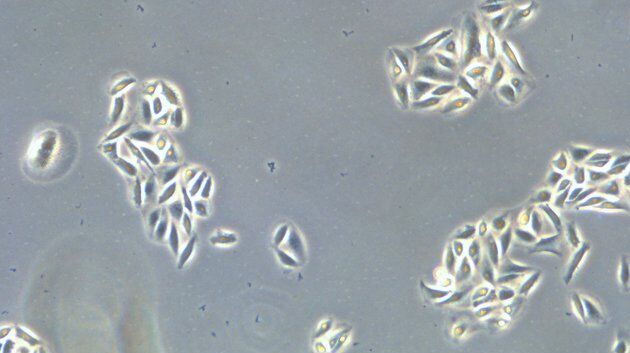
Precision and Efficiency with AI-Enhanced Cell Counting
This article describes the use of artificial intelligence (AI) for precise and efficient cell counting. Accurate cell counting is important for research with 2D cell cultures, e.g., cellular dynamics,…
Loading...
 and analyzed for confluency using AI (right).")
AI Confluency Analysis for Enhanced Precision in 2D Cell Culture
This article explains how efficient, precise confluency assessment of 2D cell culture can be done with artificial intelligence (AI). Assessing confluency, the percentage of surface area covered,…
Loading...
.")
Neuron Isolation in Spatial Context with Laser Microdissection (LMD)
After Alzheimer’s disease, Parkinson’s is the second most common progressive neurodegenerative disease. Before the first symptoms manifest, up to 70% of dopamine-releasing neurons in the mid-brain…
Loading...

How did Laser Microdissection enable Pioneering Neuroscience Research?
Dr. Marta Paterlini, a Senior Scientist at the Karolinska Institute, shares her experience of using laser microdissection (LMD) in groundbreaking research into adult human neurogenesis and offers…
Loading...

Laser Microdissection Protocols for Tissue and Cell Isolation - Download free eBook
In this Bio-protocol Selections, we present a collection of open-access, detailed methods papers using LCM to purify and isolate tissues and cells from plants, mouse embryos, cancer cells, neurons,…
Loading...

How do Cells Talk to Each Other During Neurodevelopment?
Professor Silvia Capello presents her group’s research on cellular crosstalk in neurodevelopmental disorders, using models such as cerebral organoids and assembloids.
Loading...

Workflow Solutions for Sample Preparation Methods for Material Science
This brochure presents and explains appropriate workflow solutions for the most frequently required sample preparation methods for material science samples.
Loading...
.")
Dual-View LightSheet Microscope for Large Multicellular Systems
Visualizing the dynamics of complex multicellular systems is a fundamental goal in biology. To address the challenges of live imaging over large spatiotemporal scales, Franziska Moos et. al. present…
Loading...

A Meta-cancer Analysis of the Tumor Spatial Microenvironment
Learn how clustering analysis of Cell DIVE datasets in Aivia can be used to understand tissue-specific and pan-cancer mechanisms of cancer progression
