
Biowissenschaften
Biowissenschaften
Hier können Sie Ihr Wissen, Ihre Forschungsfähigkeiten und Ihre praktischen Anwendungen der Mikroskopie in verschiedenen wissenschaftlichen Bereichen erweitern. Erfahren Sie, wie Sie präzise Visualisierung, Bildinterpretation und Forschungsfortschritte erzielen können. Hier finden Sie aufschlussreiche Informationen über fortgeschrittene Mikroskopie, Bildgebungsverfahren, Probenvorbereitung und Bildanalyse. Zu den behandelten Themen gehören Zellbiologie, Neurowissenschaften und Krebsforschung mit Schwerpunkt auf modernsten Anwendungen und Innovationen.
Filter articles
Tags
Berichtstyp
Produkte
Loading...

An Introduction to Computational Clearing
Many software packages include background subtraction algorithms to enhance the contrast of features in the image by reducing background noise. The most common methods used to remove background noise…
Loading...
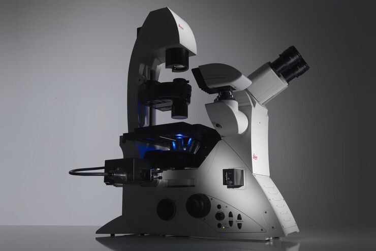
Factors to Consider When Selecting a Research Microscope
An optical microscope is often one of the central devices in a life-science research lab. It can be used for various applications which shed light on many scientific questions. Thereby the…
Loading...
Computational Clearing - Enhance 3D Specimen Imaging
This webinar is designed to clarify crucial specifications that contribute to THUNDER Imagers' transformative visualization of 3D samples and improvements within a researcher's imaging-related…
Loading...
THUNDER Imagers: High Performance, Versatility and Ease-of-Use for your Everyday Imaging Workflows
This webinar will showcase the versatility and performance of THUNDER Imagers in many different life science applications: from counting nuclei in retina sections and RNA molecules in cancer tissue…
Loading...
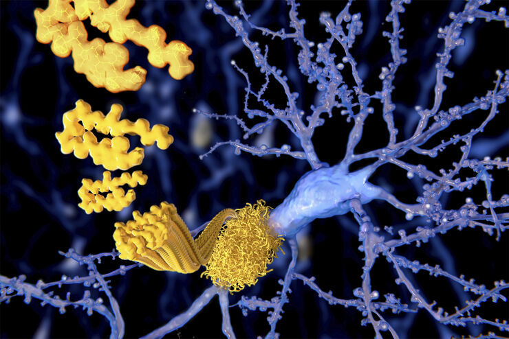
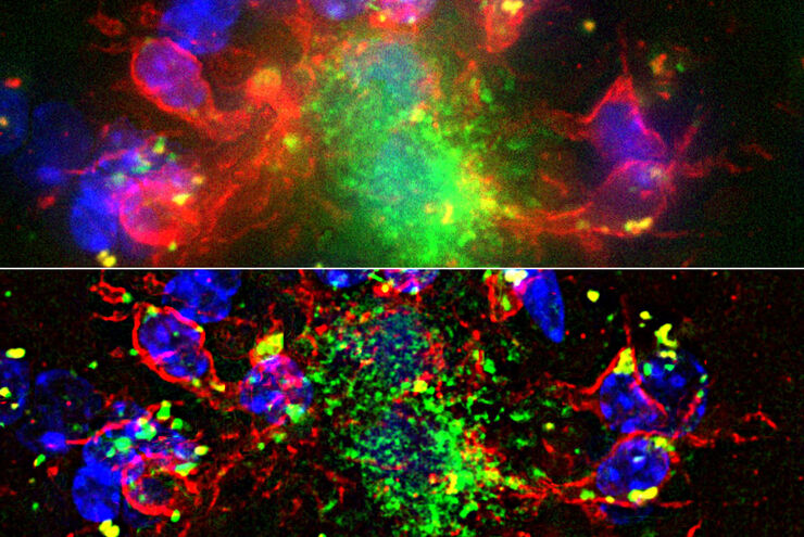
Alzheimer Plaques: fast Visualization in Thick Sections
More than 60% of all diagnosed cases of dementia are attributed to Alzheimer’s disease. Typical of this disease are histological alterations in the brain tissue. So far, there is no cure for this…
Loading...
 and YOYO 1 iodide (Nucleus).")
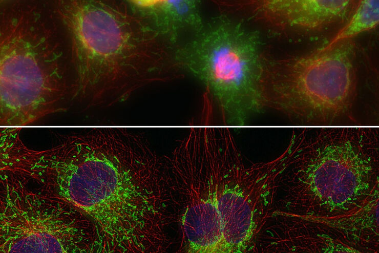
Real Time Images of 3D Specimens with Sharp Contrast Free of Haze
THUNDER Imagers deliver in real time images of 3D specimens with sharp contrast, free of the haze or out-of-focus blur typical of widefield systems. They can even image clearly places deep inside a…
