
Biowissenschaften
Biowissenschaften
Hier können Sie Ihr Wissen, Ihre Forschungsfähigkeiten und Ihre praktischen Anwendungen der Mikroskopie in verschiedenen wissenschaftlichen Bereichen erweitern. Erfahren Sie, wie Sie präzise Visualisierung, Bildinterpretation und Forschungsfortschritte erzielen können. Hier finden Sie aufschlussreiche Informationen über fortgeschrittene Mikroskopie, Bildgebungsverfahren, Probenvorbereitung und Bildanalyse. Zu den behandelten Themen gehören Zellbiologie, Neurowissenschaften und Krebsforschung mit Schwerpunkt auf modernsten Anwendungen und Innovationen.
Filter articles
Tags
Berichtstyp
Produkte
Loading...

Wie Sie Gewebeproben für die Immunfluoreszenz-Mikroskopie vorbereiten
Immunfluoreszenz (IF) ist eine leistungsfähige Methode zur Visualisierung intrazellulärer Prozesse, Bedingungen und Strukturen. IF-Präparate können mit verschiedenen Mikroskopietechniken (z. B. CLSM,…
Loading...

Fluorescent Dyes
A basic principle in fluorescence microscopy is the highly specific visualization of cellular components with the help of a fluorescent agent. This can be a fluorescent protein – for example GFP –…
Loading...

Designing your Research Study with Multiplexed IF Imaging
Multiplexed tissue analysis is a powerful technique that allows comparisons of cell-type locations and cell-type interactions within a single fixed tissue sample. It is common for researchers to ask…
Loading...

Be Confident in your Results with Cell DIVE Validated Antibodies
The Cell DIVE System includes a carefully curated list of hundreds of commercially available antibodies validated to offer optimal specificity and sensitivity in multiplexed imaging. That validation…
Loading...
 - THUNDER Imager 3D Cell Culture Influenca virus – red, cilia – green, Nuclei – blue.")
How Can Immunofluorescence Aid Virology Research?
Modern virology research has become as crucial now as ever before due to the global COVID-19 pandemic. There are many powerful technologies and assays that virologists can apply to their research into…
Loading...

Chronic Inflammation Under the Microscope
In the course of chronic inflammation certain body areas are recurrently inflamed. This goes along with many human diseases. With the help of widefield light microscopy, the underlying processes can…
Loading...
BABB Clearing and Imaging for High Resolution Confocal Microscopy
Multipohoton microscopy experiment using Leica TCS SP8 MP and Leica 20x/0.95 NA BABB immersion objective.
Understanding kidney microanatomy is key to detecting and identifying early events in kidney…
Loading...
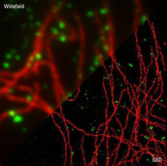
Sample Preparation for GSDIM Localization Microscopy – Protocols and Tips
The widefield super-resolution technique GSDIM (Ground State Depletion followed by individual molecule return) is a localization microscopy technique that is capable of resolving details as small as…
