
Biowissenschaften
Biowissenschaften
Hier können Sie Ihr Wissen, Ihre Forschungsfähigkeiten und Ihre praktischen Anwendungen der Mikroskopie in verschiedenen wissenschaftlichen Bereichen erweitern. Erfahren Sie, wie Sie präzise Visualisierung, Bildinterpretation und Forschungsfortschritte erzielen können. Hier finden Sie aufschlussreiche Informationen über fortgeschrittene Mikroskopie, Bildgebungsverfahren, Probenvorbereitung und Bildanalyse. Zu den behandelten Themen gehören Zellbiologie, Neurowissenschaften und Krebsforschung mit Schwerpunkt auf modernsten Anwendungen und Innovationen.
Filter articles
Tags
Berichtstyp
Produkte
Loading...

Automotive Part Verification and Development according to Specifications
Automotive part verification during the development and production of parts and components by suppliers or manufacturers is important for ensuring that specifications are met. Specifications are…
Loading...

A Guide to Darkfield Microscopes
A darkfield microscope offers a way to view the structures of many types of biological specimens in greater contrast without the need of stains.
Loading...
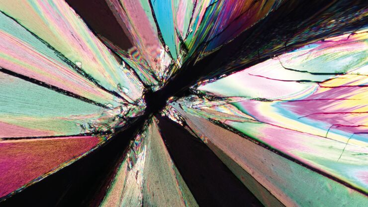
Das Prinzip der Polarisationsmikroskopie
Die Polarisationsmikroskopie wird in den Material- und Geowissenschaften routinemäßig eingesetzt, um Materialien und Mineralien anhand ihrer charakteristischen Brechungseigenschaften und Farben zu…
Loading...

Rapidly Visualizing Magnetic Domains in Steel with Kerr Microscopy
The rotation of polarized light after interaction with magnetic domains in a material, known as the Kerr effect, enables the investigation of magnetized samples with Kerr microscopy. It allows rapid…
Loading...
. With DIC users are able to visualize small height differences on the wafer surface more easily.")
6-Inch Wafer Inspection Microscope for Reliably Observing Small Height Differences
A 6-inch wafer inspection microscope with automated and reproducible DIC (differential interference contrast) imaging, no matter the skill level of users, is described in this article. Manufacturing…
Loading...

Visualizing Photoresist Residue and Organic Contamination on Wafers
As the scale of integrated circuits (ICs) on semiconductors passes below 10 nm, efficient detection of organic contamination, like photoresist residue, and defects during wafer inspection is becoming…
Loading...

Workflow Solutions for Sample Preparation Methods for Material Science
This brochure presents and explains appropriate workflow solutions for the most frequently required sample preparation methods for material science samples.
Loading...
. Das Bild wurde mit einem Digitalmikroskop DVM6 aufgenommen.")
Graterkennung während der Batterieherstellung
Erfahren Sie, wie die optische Mikroskopie zur Graterkennung an Batterieelektroden und zur Bestimmung des Schadenspotenzials eingesetzt werden kann, um eine schnelle und zuverlässige…
Loading...

Erkennung von Batteriepartikeln während des Produktionsprozesses
In diesem Artikel wird erläutert, wie die Partikelerkennung und -analyse von Batterien mit optischer Mikroskopie und Laserspektroskopie für eine schnelle, zuverlässige und kostengünstige…
