Leica Microsystems

Leica Microsystems

Leica Microsystems ist ein weltweit führender Hersteller von Mikroskopen und wissenschaftlichen Instrumenten. Im 19. Jahrhundert als Familienunternehmen gegründet, war die Geschichte des Unternehmens auf dem Weg zum Weltkonzern von beispielloser Innovation geprägt.

Die traditionell enge Zusammenarbeit mit der Wissenschaft ist der Schlüssel zur Innovationstradition von Leica Microsystems, die auf die Ideen der Anwender zurückgreift und auf deren Bedürfnisse zugeschnittene Lösungen schafft. Auf globaler Ebene ist Leica Microsystems in drei Divisionen gegliedert, die alle zu den führenden Anbietern in ihrem jeweiligen Bereich gehören: Life Science, Industrie und Medizin.

Das Unternehmen ist in über 100 Ländern mit 6 Produktionsstandorten in 5 Ländern, Vertriebs- und Serviceorganisationen in 20 Ländern und einem internationalen Händlernetz vertreten. Der Hauptsitz des Unternehmens liegt in Wetzlar, Deutschland.

https://www.leica-microsystems.com/de

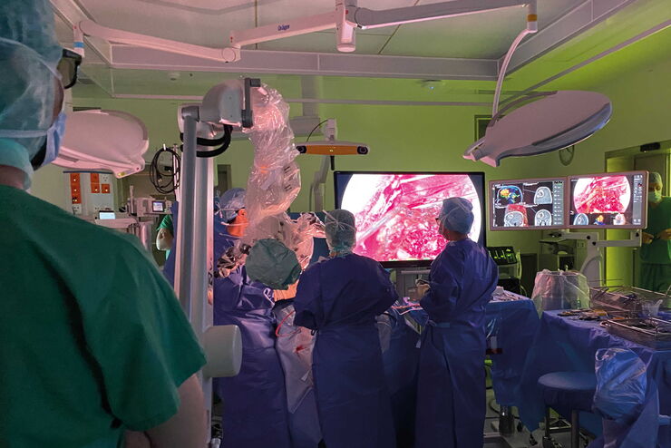
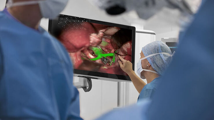
Oncological Maxillofacial Reconstructive Surgery at the Paris Saint-Joseph Hospital with Dr. Benichou. The Leica M530 OHX microscope is used by plastic surgeons in a comfortable standing up position.

Plastische und rekonstruktive Chirurgie: Warum ein Mikroskop benutzen?

Verfahren der plastischen und rekonstruktiven Chirurgie können heikel sein. Visualisierungslösungen spielen eine wichtige Rolle, da sie es ermöglichen, die Operation unter bestmöglichen Bedingungen…

Minimally Invasive Spine Surgery: Improving Precision and Accuracy with Microscopes

Spine surgery is extremely delicate and requires extensive training and experience. Innovative visualization technologies can also help achieve better outcomes allowing to see more and have a clearer…

Neurosurgery with Heads-up Display

In the following video interviews Prof. Dr. Raphael Guzman, Vice Chairman of the Department of Neurosurgery at the University Hospital in Basel, Switzerland, talks about his experience in heads-up…

Workflows and Instrumentation for Cryo-electron Microscopy

Cryo-electron microscopy is an increasingly popular modality to study the structures of macromolecular complexes and has enabled numerous new insights in cell biology. In recent years, cryo-electron…
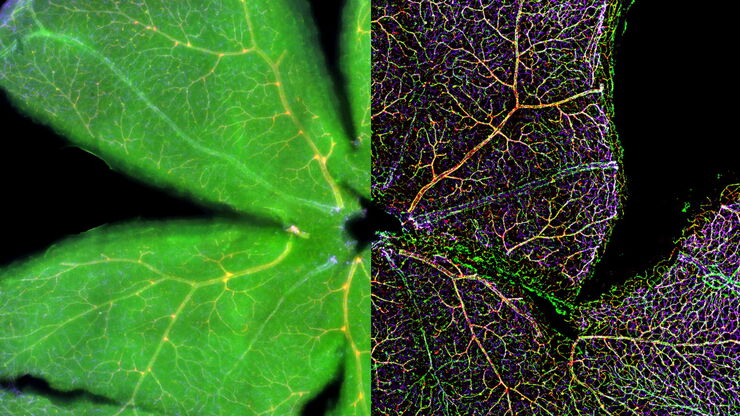
Mouse retina was fixed and stained by following reagents: anti-CD31 antibody (green): Endothelia cells, IsoB4 (red): Blood vessels, and microglia anti-GFAP antibody (blue): Astrocytes Sample courtesy by Jeremy Burton, PhD and Jiyeon Lee, PhD, Genentech Inc., South San Francisco, USA. Imaged by Olga Davydenko, PhD (Leica). Imaged with a THUNDER Imager 3D Cell Culture.

An Introduction to Computational Clearing

Many software packages include background subtraction algorithms to enhance the contrast of features in the image by reducing background noise. The most common methods used to remove background noise…

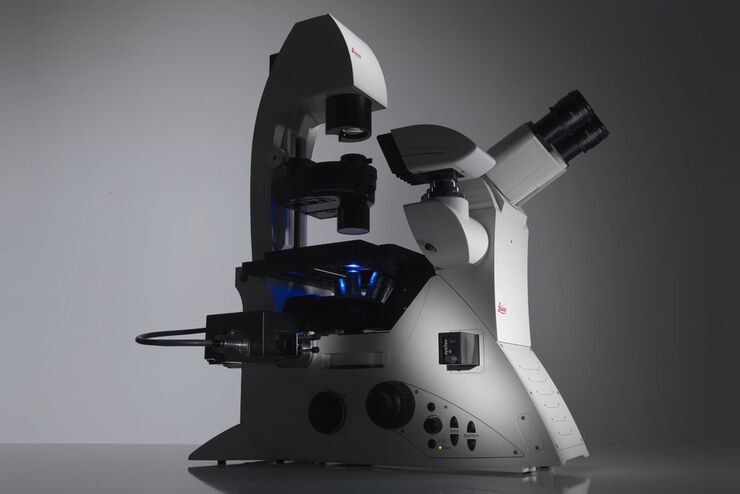
Factors to Consider When Selecting a Research Microscope

An optical microscope is often one of the central devices in a life-science research lab. It can be used for various applications which shed light on many scientific questions. Thereby the…

Challenges Faced When Manually Rating Non-Metallic Inclusions (NMIs) to Determine Steel Quality

Rapid, accurate, and reliable rating of non-metallic inclusions (NMIs) is instrumental for the determination of steel quality. This article describes the challenges that arise from manual NMI rating,…
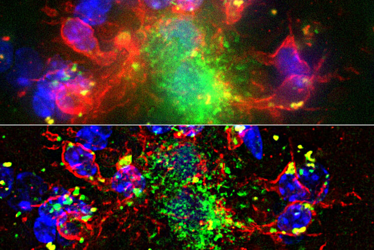
Influenca in lung epithelial cells (porcine) - THUNDER Imager 3D Cell Culture Influenca virus – red, cilia – green, Nuclei – blue.

How Can Immunofluorescence Aid Virology Research?

Modern virology research has become as crucial now as ever before due to the global COVID-19 pandemic. There are many powerful technologies and assays that virologists can apply to their research into…

Augmented Reality (AR) Fluorescence Image Gallery

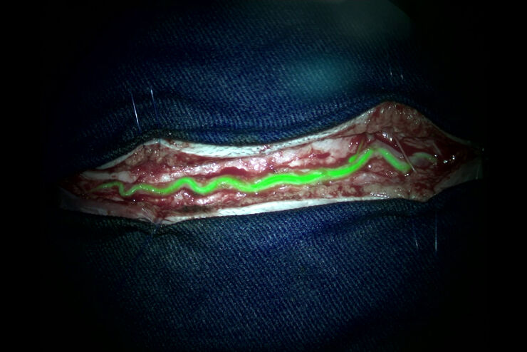
Building on a decade of leadership in fluorescence imaging technology, GLOW800 AR fluorescence is the first of many modalities based on the proprietary GLOW AR platform. The sophisticated imaging…

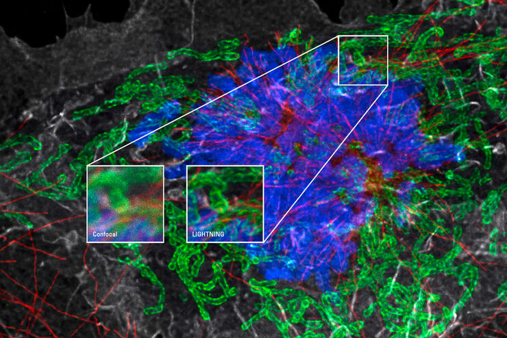
Mit LIGHTNING das Maximum an Informationen aus Ihrer Probe erhalten

LIGHTNING ist ein adaptiver Prozess zur Extraktion von Bildinformationen, bei dem vollautomatisch anderweitig nicht sichtbare Strukturen und feine Details sichtbar gemacht werden. Im Gegensatz zu…

Advanced Techniques in Cataract and Refractive Surgery

In this webinar Dr. Thompson and Dr. Moshirfar will explain how Leica microscopes aid in procedures such as Centration of Multifocal IOLs and corneal inlays such as Kamra and Lenticular Grafts used in…

Clinical Uses in Cerebrovascular and Skull Base Neurosurgery

In this webinar Dr. Bendok and Dr. Morcos explain how Augmented Reality and Fluorescence can enhance visualization and support surgical decision making. They present first-hand experience of the GLOW…

Industrial Microscopy: Digital imaging and the Leica DVM6

This webinar will discuss digital microscopy and Leica’s digital DVM6 microscope. We will navigate the difference between optical and digital magnification, explain the differences in optics, and…

Reasons Why There is Growing Need for Fast and Reliable Steel Quality Rating Solutions

Steel quality is critical for the manufacturing of high-quality components and products. Fast, reliable, and accurate detection and classification of inclusions has become essential for both component…

Inverted Microscopes for Grain Size Analysis: Three Factors to Consider

Microscopic steel grain size analysis is useful in determining the quality of steel alloys for a given purpose such as building bridges vs railroad rails. This webinar will describe the preparation of…

Crystal Clear Cryo Light-microscopy Images

This article describes how computational clearing of cryo light microscopy images improves the identification of cellular targets for cryo electron-microscopy.

Depth Profiling and Layer Analysis for Inspection of Materials with a 2-In-1 Solution Combining Optical Microscopy and Laser Spectroscopy

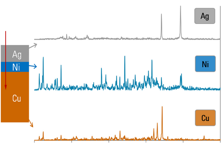
In addition to simultaneous visual and chemical inspection, a 2-methods-in-1 materials analysis solution, which combines optical microscopy and laser induced breakdown spectroscopy (LIBS), can also be…

Microscopy in Virology

The coronavirus SARS-CoV-2, causing the Covid-19 disease effects our world in all aspects. Research to find immunization and treatment methods, in other words to fight this virus, gained highest…

Surgical Microscopes: Key Factors for OR Nurses

Operating room (OR) nurses are vital to the surgery process. An OR Nurse Manager explains the key surgical microscope features to facilitate their work.

Top Issues Related to Standards for Rating Non-Metallic Inclusions in Steel

Supplying components and products made of steel to users worldwide can require that a single batch be compliant with multiple steel quality standards. This user demand creates significant challenges…
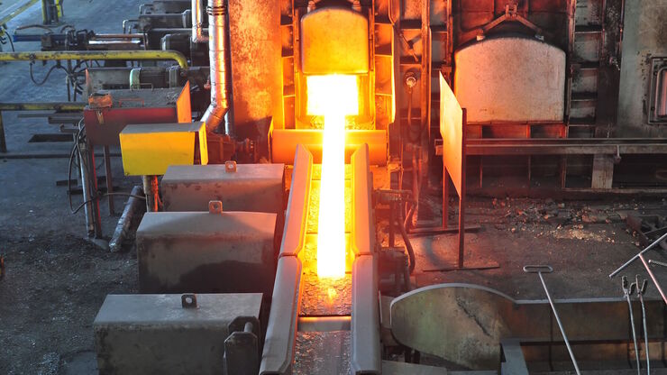
Steelworks

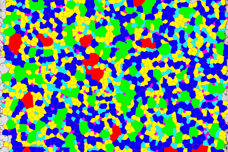
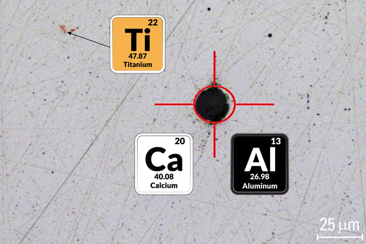
Analyzing Non-metallic Inclusions in Steel

Oftentimes we find ourselves caught up in tedious analyses by reticle and comparison chart, time-consuming double-evaluation according to several standards or subjective inspection results with a bias…
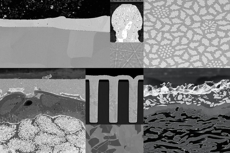
Printed Circuit Board (PCB)

Performing Elemental Analysis down to the Micro Scale

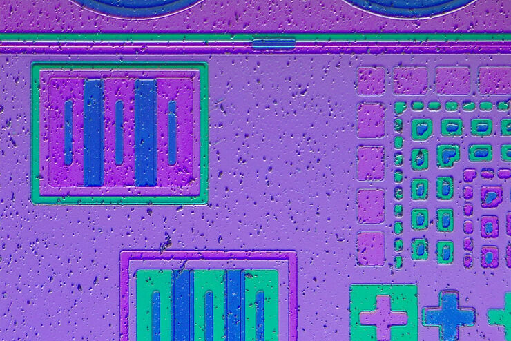
If you work in electronic component analysis, you will be familiar with the many challenges posed. Whether you are identifying metallic particles or checking product authenticity, it’s important to…

Introduction to Ion Beam Etching with the EM TIC 3X

In this article you can learn how to optimize the preparation quality of your samples by using the ion beam etching method with the EM TIC 3X ion beam milling machine. A short introduction of the…

Computational Clearing - Enhance 3D Specimen Imaging

This webinar is designed to clarify crucial specifications that contribute to THUNDER Imagers' transformative visualization of 3D samples and improvements within a researcher's imaging-related…

Visual and Chemical Analysis of Steel Microstructure: Faster Rating of Steel Quality

Simultaneous visual and chemical analysis of steel non-metallic inclusions with a 2-methods-in-1 solution, using optical microscopy and laser induced breakdown spectroscopy (LIBS), is described in…

Explore Innovative Techniques to Separate Fluorophores with Overlapping Spectra

In this article we explore several strategies you can take to improve the separation of fluorophores and increase the number of fluorescent probes you can distinguish in your sample.

STELLARIS White Light Lasers

When it comes to choosing fluorescent probes for your multi-color experiments, you shouldn’t have to compromise. Now you can advance beyond conventional excitation sources that limit your fluorophore…

Workflow Solutions for Sample Preparation Methods for Material Science

This brochure presents and explains appropriate workflow solutions for the most frequently required sample preparation methods for material science samples.

Rate the Quality of Your Steel: Free Webinar and Report

This webinar and report describe optimal microscopy solutions for rating steel quality in terms of non-metallic inclusions and reviews the various international and regional standards concerning…

The Power HyD Detector Family

Powerful photon counting detectors on the STELLARIS confocal platform provide improved photon counting, ultra-sensitive imaging and more color options in the NIR spectrum.
Scroll to top