Kontaktieren Sie uns

Leica Microsystems

Leica Microsystems

Leica Microsystems ist ein weltweit führender Hersteller von Mikroskopen und wissenschaftlichen Instrumenten. Im 19. Jahrhundert als Familienunternehmen gegründet, war die Geschichte des Unternehmens auf dem Weg zum Weltkonzern von beispielloser Innovation geprägt.

Die traditionell enge Zusammenarbeit mit der Wissenschaft ist der Schlüssel zur Innovationstradition von Leica Microsystems, die auf die Ideen der Anwender zurückgreift und auf deren Bedürfnisse zugeschnittene Lösungen schafft. Auf globaler Ebene ist Leica Microsystems in drei Divisionen gegliedert, die alle zu den führenden Anbietern in ihrem jeweiligen Bereich gehören: Life Science, Industrie und Medizin.

Das Unternehmen ist in über 100 Ländern mit 6 Produktionsstandorten in 5 Ländern, Vertriebs- und Serviceorganisationen in 20 Ländern und einem internationalen Händlernetz vertreten. Der Hauptsitz des Unternehmens liegt in Wetzlar, Deutschland.

https://www.leica-microsystems.com/de

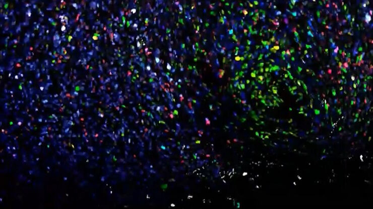
[Translate to German:] Co-detection of 10 extracellular matrix proteins and 3 topographical tissue landmarks by multiplex immunostaining within a single high-grade fibrous hotspot from a human hepatocellular carcinoma

In-situ-Identifizierung von Krebsstammzellnischen im Leberzellkarzinom

Die Multiplex-Bildgebung spielt eine entscheidende Rolle bei der Krebsforschung, da sie es Wissenschaftlern ermöglicht, tief in die komplexe Mikroumgebung von Tumoren einzudringen und einzigartige…
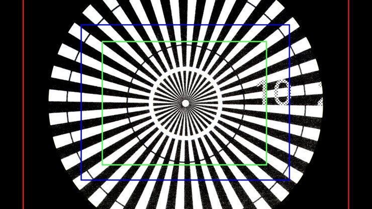
Image of a Siemens star, where the diameter of the 1st black line circle is 10 mm and the 2nd is 20 mm, taken via an eyepiece of a M205 A stereo microscope. The rectangles represent the field of view (FOV) of a Leica digital camera when installed with various C-mounts (red 0.32x, blue 0.5x, green 0.63x).

Understanding Clearly the Magnification of Microscopy

To help users better understand the magnification of microscopy and how to determine the useful range of magnification values for digital microscopes, this article provides helpful guidelines.
Fluorescence microscopy image of liver tissue where DNA in the nuclei are stained with Feulgen-pararosanilin and visualized with transmitted green light.

Epi-Illumination Fluorescence and Reflection-Contrast Microscopy

This article discusses the development of epi-illumination and reflection contrast for fluorescence microscopy concerning life-science applications. Much was done by the Ploem research group…
Esophageal tissue with a squamous cell carcinoma labelled with the 4 biomarkers PanCk, DAPI, NaKATPase, and Vimentin.

Discover how Multiplexed Bioimaging can Advance Cancer Research

Explore multiplexing with up to 60 biomarkers, enabling advanced tumor imaging approaches to gather precise, spatially-resolved single-cell data that helps enhance cancer research and clinical…
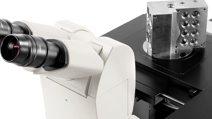
Material sample with a large height, size, and weight being observed with an inverted microscope.

Five Inverted-Microscope Advantages for Industrial Applications

With inverted microscopes, you look at samples from below since their optics are placed under the sample, with upright microscopes you look at samples from above. Traditionally, inverted microscopes…
Application example of hyperspectral imaging

Potential of Multiplex Confocal Imaging for Cancer Research and Immunology

Explore the new frontiers of multi-color fluorescent imaging: from image acquisition to analysis
Serious Game in Intraoperative Neurosurgery. Image courtesy of Dr. Lucas Troude.

Enhancing Neurosurgery Teaching

Learn about the Serious Game in Intraoperative Neurosurgery and how it supports neurosurgical teaching and the acquisition of decision-making skills.
Acrylonitrile butadiene styrene (ABS) stained with osmium tetroxide (OsO4), sectioned with a DIATOME diamond knife at room temperature, and then imaged with HAADF TEM.

Ultramicrotomy Techniques for Materials Sectioning

Learn about ultramicrotomy for materials sectioning when investigating polymers and brittle materials with transmission (TEM) or scanning electron microscopy (SEM) or atomic force microscopy.
Single cell datasets

Exploring Subcellular Spatial Phenotypes with SPARCS

Discover spatially resolved CRISPR screening (SPARCS), a platform for microscopy-based genetic screening for spatial subcellular phenotypes at the human genome scale.
Molecular structure of the green fluorescent protein (GFP)

Introduction to Fluorescent Proteins

Overview of fluorescent proteins (FPs) from, red (RFP) to green (GFP) and blue (BFP), with a table showing their relevant spectral characteristics.
Caption: Collaboration is key Picture copyright: Donenko Oleksii, Shutterstock

Launching a Neurosurgical Department with Limited Resources

Learn about Dr. Claire Karekezi’s journey and experience launching a neurosurgical department within the Rwanda Military Hospital with limited resources.

Coherent Raman Scattering Microscopy Publication List

CRS (Coherent Raman Scattering) microscopy is an umbrella term for label-free methods that image biological structures by exploiting the characteristic, intrinsic vibrational contrast of their…
Image of an onion flake taken with a basic Leica compound microscope after it was tested for resistance to fungus and mold growth following part 11 of the ISO 9022 standard.

ISO 9022 Standard Part 11 - Testing Microscopes with Severe Conditions

This article describes a test to determine the robustness of Leica microscopes to mold and fungus growth. The test follows the specifications of the ISO 9022 part 11 standard for optical instruments.
Image of an integrated-circuit (IC) chip cross section acquired at higher magnification showing a region of interest.

Structural and Chemical Analysis of IC-Chip Cross Sections

This article shows how electronic IC-chip cross sections can be efficiently and reliably prepared and then analyzed, both visually and chemically at the microscale, with the EM TXP and DM6 M LIBS…
EBSD grain size distribution of the cross section of a gold wire within a silicon matrix from inside a CPU (central processing unit of a computer). The grains are highlighted with arbitrary colors.

High-Quality EBSD Sample Preparation

This article describes a method for EBSD sample preparation of challenging materials. The high-quality samples required for electron backscatter diffraction are prepared with broad ion-beam milling.
Augmented Reality fluorescence supports each step of neurovascular surgery procedures. Image courtesy of Dr. Christof Renner.

Use of AR Fluorescence in Neurovascular Surgery

Learn about the use of GLOW800 Augmented Reality in neurovascular surgery through clinical cases and videos, including aneurysm and tumor resection cases.
Ausgewachsene hermaphroditische Gonaden von C. elegans, aufgenommen mit THUNDER Imager. Färbung: blau - DAPI (Zellkern), grün - SP56 (Spermien), rot - RME-2 (Eizelle), magenta - PGL-1 (RNA + Proteingranula). Bild mit freundlicher Genehmigung von Prof. Dr. Christian Eckmann, Martin-Luther-Universität, Halle, Deutschland.

Life-Science-Forschung: Welche Mikroskopkamera ist die richtige für Sie?

Wie Sie entscheiden, welche Kamera für Ihre Life-Science-Mikroskopie-Experimente die Richtige ist. Welche Kamera von Leica Microsystems ist für Sie am besten geeignet?
Pancreatic Ductal Adenocarcinoma with 11 Apoptosis biomarkers shown – BAK, BAX, BCL2, BCLXL, Caspase9, CIAP1, NaKATPase, PCK26, SMAC, Vimentin, and XIAP.

Multiplexing with Luke Gammon: Advance your Spatial Biology Research

Learn how multiplexing imaging and spatial biology can help researchers better understand complex biological systems. In this interview, Dr. Gammon and Dr. Pointu of Leica Microsystems discuss pain…
Scroll to top