Kontaktieren Sie uns

Leica Microsystems

Leica Microsystems

Leica Microsystems ist ein weltweit führender Hersteller von Mikroskopen und wissenschaftlichen Instrumenten. Im 19. Jahrhundert als Familienunternehmen gegründet, war die Geschichte des Unternehmens auf dem Weg zum Weltkonzern von beispielloser Innovation geprägt.

Die traditionell enge Zusammenarbeit mit der Wissenschaft ist der Schlüssel zur Innovationstradition von Leica Microsystems, die auf die Ideen der Anwender zurückgreift und auf deren Bedürfnisse zugeschnittene Lösungen schafft. Auf globaler Ebene ist Leica Microsystems in drei Divisionen gegliedert, die alle zu den führenden Anbietern in ihrem jeweiligen Bereich gehören: Life Science, Industrie und Medizin.

Das Unternehmen ist in über 100 Ländern mit 6 Produktionsstandorten in 5 Ländern, Vertriebs- und Serviceorganisationen in 20 Ländern und einem internationalen Händlernetz vertreten. Der Hauptsitz des Unternehmens liegt in Wetzlar, Deutschland.

https://www.leica-microsystems.com/de

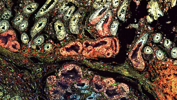
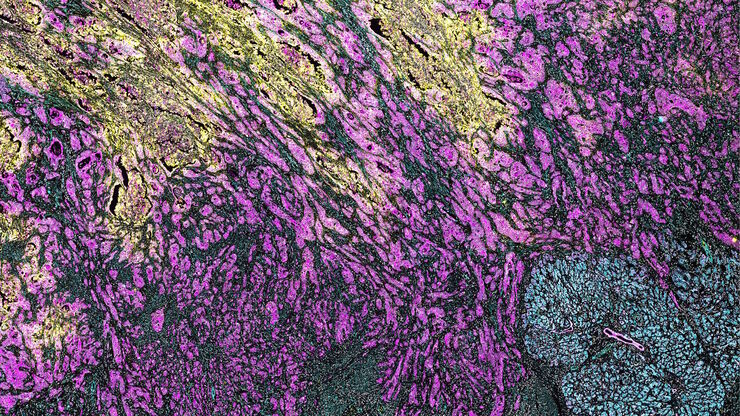
[Translate to German:] Multi-tissue array with 4 markers shown including DAPI, NaKATPase, PanCk, and Vimentin.

Räumliche Biologie: Erwägung neuer Wege

Räumliche Biologie: Forschung zu Anordnung und Interaktion von Molekülen, Zellen und Geweben in ihrem nativen räumlichen Kontext
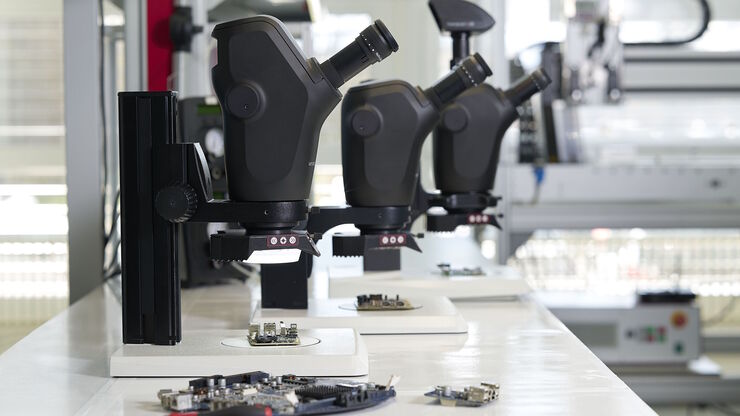
[Translate to German:] Stereo microscopes are often considered the workhorses of laboratories and production sites.

Wichtige Faktoren, die Sie bei der Auswahl eines Stereomikroskops berücksichtigen sollten

Stereomikroskope zeichnen sich durch ihre Fähigkeit aus, einen 3D-Eindruck der Probe zu erzeugen. Daher eignen sie sich besonders gut für Inspektion und Nacharbeit, Qualitätskontrolle, Forschung und…
Brain organoid section (DAPI) acquired using THUNDER Imager Live Cell. Image courtesy of Janina Kaspar and Irene Santisteban, Schäfer Lab, TUM.

Imaging Organoid Models to Investigate Brain Health

Imaging human brain organoid models to study the phenotypes of specialized brain cells called microglia, and the potential applications of these organoid models in health and disease.
Geringe Vergrößerung DVM6-Bild eines Teils der PCBA-Probe.

Schnelle und zuverlässige Untersuchung von Leiterplatten und Leiterplattenbaugruppen mittels Digitalmikroskopie

Digitalmikroskope bieten Anwendern eine bequeme und schnelle Möglichkeit zur Erfassung hochwertiger, zuverlässiger Bilddaten und zur schnellen Inspektion und Analyse von Leiterplatten (PCBs) und…
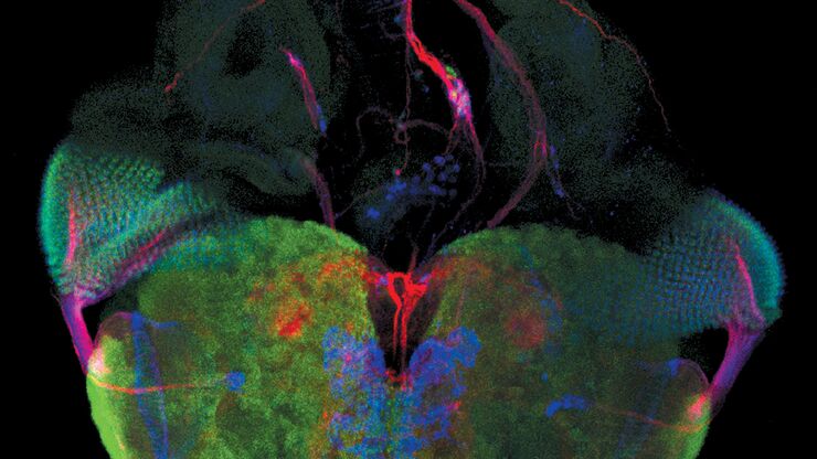
In vivo imaging of a mouse pial and cortical vasculature through a glass window (ROSAmT/mG::Pdgfb-CreERT2 mouse meningeal and cortical visualization following tamoxifen induction and craniotomy). Courtesy: Thomas Mathivet, PhD

Windows on Neurovascular Pathologies

Discover how innate immunity can sustain deleterious effects following neurovascular pathologies and the technological developments enabling longitudinal studies into these events.
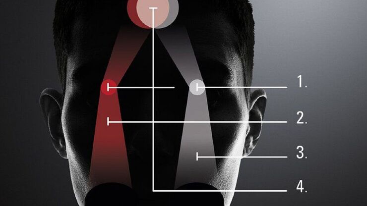
The principle of the FusionOptics technology:  Of the two separate beam paths (1), one provides depth of field (2) and the other high resolution (3). In the brain, the two images of the sample are merged into a single, optimal 3D image (4).

What is the FusionOptics Technology?

Leica stereo microscopes with FusionOptics provide optimal 3D perception. The brain merges two images, one with large depth of field and the other with high resolution, into one 3D image.
Lifetime-based multiplexing in live cells using TauSeparation. Mammalian cells expressing LifeAct-GFP (ibidi GmbH) and labelled with MitoTracker Green. Acquisition with one detector, intensity information shown in grey. The two markers can be separated using lifetime information: LifeAct-GFP (cyan), MitoTracker Green (magenta). Image acquired with STELLARIS 5.

The Power of Reproducibility, Collaboration and New Imaging Technologies

In this webinar you willl learn what impacts reproducibility in microscopy, what resources and initiatives there are to improve education and rigor and reproducibility in microscopy and how…
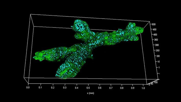
Branched organoid growing in collagen where the Nuclei are labeled blue. To detect the mechanosignaling process, the YAP1 is labeled green.

Examining Developmental Processes In Cancer Organoids

Interview: Prof. Bausch and Dr. Pastucha, Technical University of Munich, discuss using microscopy to study development of organoids, stem cells, and other relevant disease models for biomedical…
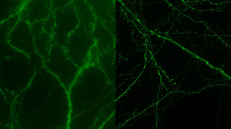
Mouse cortical neurons. Transgenic GFP (green). Image courtesy of Prof. Hui Guo, School of Life Sciences, Central South University, China

How Microscopy Helps the Study of Mechanoceptive and Synaptic Pathways

In this podcast, Dr Langenhan explains how microscopy helps his team to study mechanoceptive and synaptic pathways, their challenges, and how they overcome them.
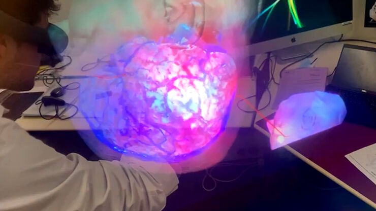
Advanced technologies support neurosurgical teaching. Image courtesy of Dr. Florian Bernard.

3D, AR & VR for Teaching in Neurosurgery

Discover the evolution of neurosurgical teaching and how 3D, Augmented Reality and Virtual Reality can help better learn anatomy and acquire surgical skills.
[Translate to German:] Cell counts for each biomarker were divided by total number of cells to give a percentage of biomarker positive cells out of total cells for each biomarker.

Methoden zur Verbesserung der Reproduzierbarkeit in der räumlichen Biologieforschung

Durch den Einsatz von Automatisierung, hochwertigen Antikörpern und einem bewährten Multiplex-Imaging-Workflow ist Cell DIVE in der Lage, reproduzierbare Ergebnisse zu liefern. In diesem Poster…
THY1-EGFP labeled neurons in mouse brain processed using the PEGASOS 2 tissue clearing method, imaged on a Leica confocal microscope. Neurons were traced using Aivia’s 3D Neuron Analysis – FL recipe. Image credit: Hu Zhao, Chinese Institute for Brain Research.

Unlocking Insights in Complex and Dense Neuron Images Guided by AI

The latest advancement in Aivia AI image analysis software provides improved soma detection, additional flexibility in neuron tracing, 3D relational measurement including Sholl analysis and more.
PDAC Multiplexed imaging of CST panels enables an examination of immune cell components in pancreatic ductal adenocarcinoma (IPDAC) tissue on a single slide.

Characterizing tumor environment to reveal insights and spatial resolution

Antibodies from Cell Signaling Technology are validated for use with the Cell DIVE multiplexing workflow and used to probe cell lineages in the tumor microenvironment
Microscopy for neuroscience research

What are the Challenges in Neuroscience Microscopy?

eBook outlining the visualization of the nervous system using different types of microscopy techniques and methods to address questions in neuroscience.
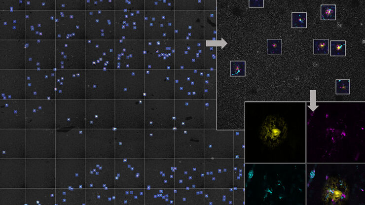
AI-based workflow for fast rare event detection in living biological samples using Autonomous Microscopy powered by Aivia

AI Microscopy Enables the Efficient Detection of Rare Events

Localization and selective imaging of rare events is key for the investigation of many processes in biological samples. Yet, due to time constraints and complexity, some experiments are not feasible…

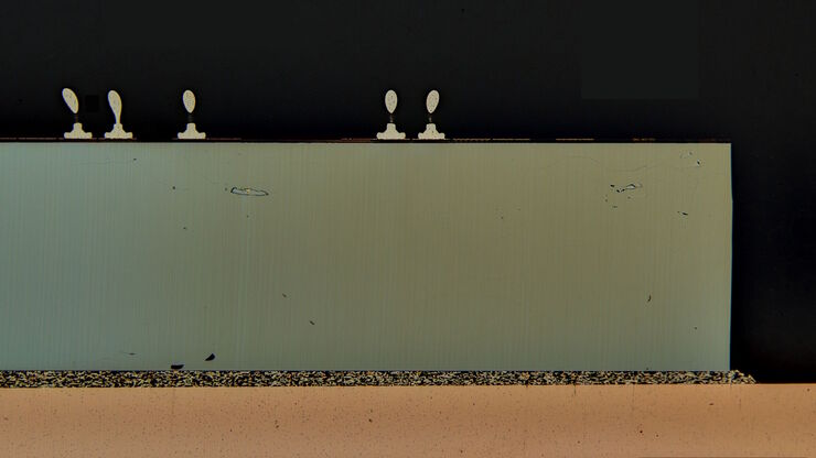
How to Prepare and Analyse Battery Samples with Electron Microscopy

This workshop covers the sample preparation process for lithium and novel battery sample analysis, as well as other semiconductor samples requiring high-resolution cross-section imaging.
Krebszellen

Die Rolle des Eisenstoffwechsels bei der Krebsentwicklung

Der Eisenstoffwechsel spielt eine Rolle bei der Entstehung und dem Fortschreiten von Krebs und beeinflusst die Immunreaktion. Zu verstehen, wie Eisen Krebs und das Immunsystem beeinflusst, kann die…
Pancreatic Ductal Adenocarcinoma with 5 biomarkers shown – SMA, PanCK PCK26, PanCK AE1, Vimentin, and Glut1.

Dig Deeper Into the Complexities of Pancreatic Cancer with Multiplex Imaging

Cell DIVE is an iterative staining workflow for multiplexed imaging that unveils biological pathways to dig deeper into the complexities of pancreatic cancer.
Scroll to top