Philipps Universität Marburg, Institut für Zytobiologie and Zytopathologie, Deutschland

Philipps Universität Marburg, Institut für Zytobiologie and Zytopathologie, Deutschland

Das Institut ist in den Fachbereich Medizin der Philipps-Universität Marburg integriert. Die Mitglieder betreiben Grundlagenforschung in Zellbiologie und biologischer Chemie. Das Institut ist auch für die Ausbildung von Studenten der Medizin, Zahnmedizin und Humanbiologie zuständig. Zentrale Forschungsprojekte befassen sich mit molekularen Mechanismen bei der Biogenese von Zellorganellen. Dabei ist das Verständnis von Mutationen der beteiligten Proteine von besonderem Interesse, da diese Krankheiten verursachen. Die Forschung erfolgt hauptsächlich an den Modellorganismen Hefe, Maus und Ratte. Weiterhin werden verschiedene Zellkultursysteme von Säugetieren verwendet. Das Methodenspektrum in den einzelnen Projekten umfasst zellbiologische, biochemische, immunhistochemische, molekularbiologische und genetische Techniken. 

Zentrale Forschungsgebiete sind:

  • Biogenese von Eisen-Schwefel-Proteinen in den Mitochondrien und im Zytosol
  • Molekulare Grundlagen der neurodegenerativen Erkrankung Friedreich'sche Ataxie
  • Mechanismus und Regulation des mitochondrialen Eisen(III)-Transports
  • Reifung und Sortierung von Proteinen in polarisierten Epithelzellen
  • Funktion von lysosomalen Proteinen
  • Funktion des Mys-bindenden Proteins Miz-1 in Keratinozyten
http://www.uni-marburg.de/fb20/cyto/

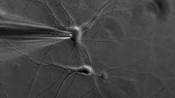
Patch pipette touching a murine hippocampal neuron. Image courtesy of A. Aguado, Ruhr University Bochum, Germany.

What is the Patch-Clamp Technique?

This article gives an introduction to the patch-clamp technique and how it is used to study the physiology of ion channels for neuroscience and other life-science fields.
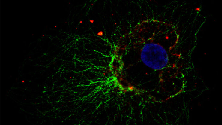
Zelluläres Mikrotubuli-Netzwerk in einer Fibroblastenzelle.

Wie Sie Gewebeproben für die Immunfluoreszenz-Mikroskopie vorbereiten

Immunfluoreszenz (IF) ist eine leistungsfähige Methode zur Visualisierung intrazellulärer Prozesse, Bedingungen und Strukturen. IF-Präparate können mit verschiedenen Mikroskopietechniken (z. B. CLSM,…
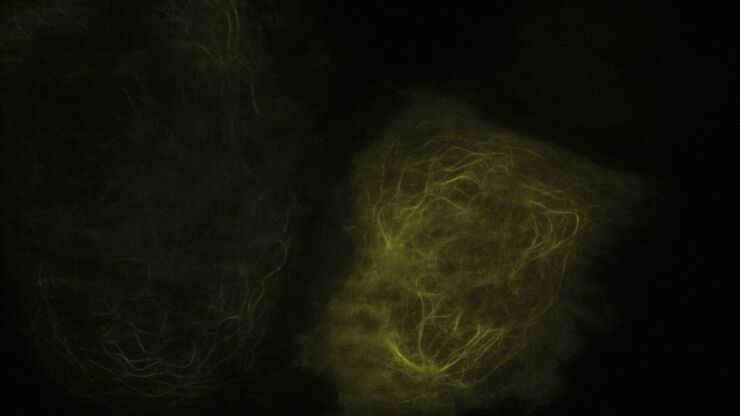
TIRF Image of Tubulin, YFP, penetration depth: 120 mm

Applications of TIRF Microscopy in Life Science Research

The special feature of TIRF microscopy is the employment of an evanescent field for fluorophore excitation. Unlike standard widefield fluorescence illumination procedures with arc lamps, LEDs or…
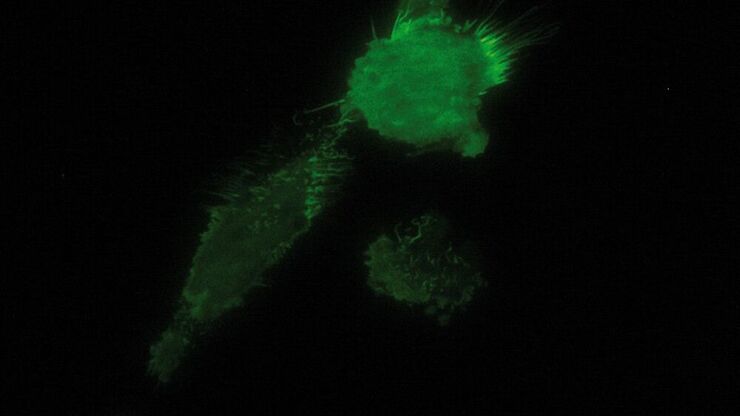
TIRF image of brest carcinoma tumor cells expressing GFP tagged cell adhesion Molecule CD44 that is expressed on the cell membrane, imagined in TIRF.

Total Internal Reflection Fluorescence (TIRF) Microscopy

Total internal reflection fluorescence (TIRF) is a special technique in fluorescence microscopy developed by Daniel Axelrod at the University of Michigan, Ann Arbor in the early 1980s. TIRF microscopy…

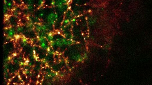
Super-Resolution GSDIM Microscopy

The nanoscopic technique GSDIM (ground state depletion microscopy followed by individual molecule return) provides a detailed image of the spatial arrangement of proteins and other biomolecules within…
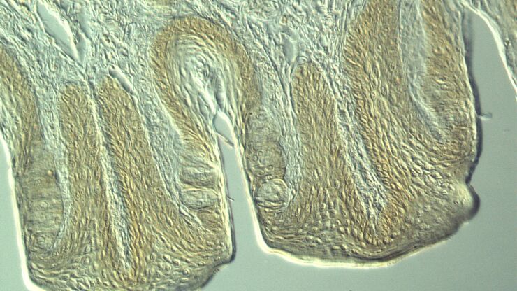
Section taste buds rabbit, differential interference contrast microscope

Optical Contrast Methods

Optical contrast methods give the potential to easily examine living and colorless specimens. Different microscopic techniques aim to change phase shifts caused by the interaction of light with the…
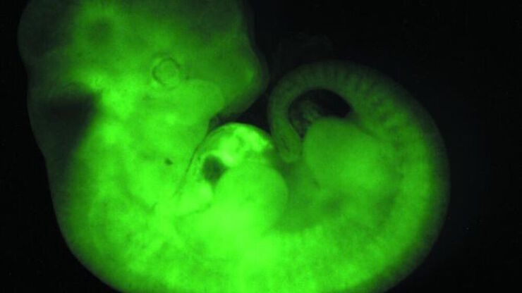
Transgenic Mouse Embryo, GFP

Fluorescence in Microscopy

Fluorescence microscopy is a special form of light microscopy. It uses the ability of fluorochromes to emit light after being excited with light of a certain wavelength. Proteins of interest can be…
Scroll to top