
Science Lab
Science Lab
Willkommen auf dem Wissensportal von Leica Microsystems. Hier finden Sie wissenschaftliches Forschungs- und Lehrmaterial rund um das Thema Mikroskopie. Das Portal unterstützt Anfänger, erfahrene Praktiker und Wissenschaftler gleichermaßen bei ihrer täglichen Arbeit und ihren Experimenten. Erkunden Sie interaktive Tutorials und Anwendungshinweise, entdecken Sie die Grundlagen der Mikroskopie ebenso wie High-End-Technologien. Werden Sie Teil der Science Lab Community und teilen Sie Ihr Fachwissen.
Filter articles
Tags
Berichtstyp
Produkte
Loading...

Imaging and Analyzing Zebrafish, Medaka, and Xenopus
Discover how to image and analyze zebrafish, medaka, and Xenopus frog model organisms efficiently with a microscope for developmental biology applications from this article.
Loading...

Bacteria Protocol - Critical Point Drying of E. coli for SEM
Application Note for Leica EM CPD300 - Critical point drying of E. coli with subsequent platinum / palladium coating and SEM analysis. Sample was inserted into a filter disc (Pore size: 16 - 40 μm)…
Loading...
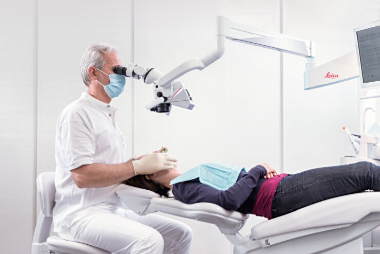
Erfolgreiche endodontische Behandlung mit Dental-Operationsmikroskopen
In der Endodontie hängt die zielgerichtete Behandlung nicht nur von den technischen Fähigkeiten und dem Wissen des Zahnarztes ab. Es kommt auch auf die klare, detaillierte Visualisierung des…
Loading...

Investigating Fruit Flies (Drosophila melanogaster)
Learn how to image and investigate Drosophila fruit fly model organisms efficiently with a microscope for developmental biology applications from this article.
Loading...

Studying Caenorhabditis elegans (C. elegans)
Find out how you can image and study C. elegans roundworm model organisms efficiently with a microscope for developmental biology applications from this article.
Loading...
BABB Clearing and Imaging for High Resolution Confocal Microscopy
Multipohoton microscopy experiment using Leica TCS SP8 MP and Leica 20x/0.95 NA BABB immersion objective.
Understanding kidney microanatomy is key to detecting and identifying early events in kidney…
Loading...
")
Epoxy Resin Embedding of Animal and Human Tissues for Pathological Diagnosis and Research
Application Note for Leica EM AMW - The tissues were fixed in the modified Karnovsky fixative generally by immersion overnight (at minimum 4h at room temperature). Then pieces of approx. 1mm3 were cut…
Loading...
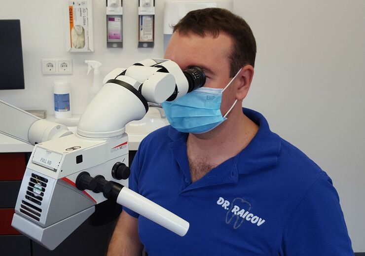
Das Dentalmikroskop in der Endodontie
In der Zahnmedizin unterstützt ein heller, vergrößerter Blick in tiefe Kavitäten die detaillierte Diagnose und präzise Therapie, insbesondere im Bereich der Endodontie. In diesem Interview erklärt Dr.…
Loading...

Human Blood Cells Protocol
Application Note for Leica EM CPD300 - Life Science Research. Species: Human (Homo sapiens)
Critical point drying of human blood with subsequent platinum / palladium coating and SEM analysis.
