
Medizinische Fachgebiete
Medizinische Fachgebiete
Entdecken Sie eine umfassende Sammlung wissenschaftlicher und klinischer Ressourcen, die speziell für Ärzte im Gesundheitswesen entwickelt wurden, darunter Berichte von Kollegen, klinische Fallstudien und Symposien. Speziell für Neurochirurgen, Augenärzte, plastische und rekonstruktive Chirurgen, HNO-Ärzte und Zahnärzte. Diese Sammlung präsentiert die neuesten Fortschritte in der chirurgischen Mikroskopie. Entdecken Sie, wie modernste chirurgische Technologien wie AR-Fluoreszenz, 3D-Visualisierung und intraoperative OCT-Bildgebung eine sichere Entscheidungsfindung und Präzision bei komplexen Eingriffen ermöglichen.
Filter articles
Tags
Produkte
Loading...

Tissue Image Gallery
Visual analysis of animal and human tissues is critical to understand complex diseases such as cancer or neurodegeneration. From basic immunohistochemistry to intravital imaging, confocal microscopy…
Loading...
 and astrocytes (green) in a cortical spheroid derived from human induced pluripotent stem cells.")
Neuroscience Images
Neuroscience commonly uses microscopy to study the nervous system’s function and understand neurodegenerative diseases.
Loading...
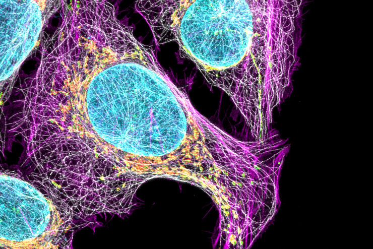
Multicolor Image Gallery
Fluorescence multicolor microscopy, which is one aspect of multiplex imaging, allows for the observation and analysis of multiple elements within the same sample – each tagged with a different…
Loading...

Cancer Research Image Gallery
Fluorescence microscopy allows the study of changes occurring in tissue and cells during cancer development and progression. Techniques such as live cell imaging are critical to understand cancer…
Loading...
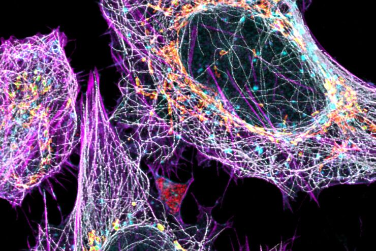
Cell Biology Image Gallery
Cell biology studies the structure, function and behavior of cells, including cell metabolism, cell cycle, and cell signaling. Fluorescence microscopes are an integral part of a cell biologist…
Loading...

Developmental Biology Image Gallery
Developmental biology explores the development of complex organisms from the embryo to adulthood to understand in detail the origins of disease. This category of the gallery shows images about…
Loading...

Vom Licht zur Erleuchtung: Sensoren und Messverfahren in der konfokalen Mikroskopie
In diesem Beitrag werden die wichtigsten Sensoren kurz vorgestellt, die in der konfokalen Mikroskopie verwendet werden. Mit konfokaler Mikroskopie ist hier „True Confo-cal Scanning“ gemeint, also das…
