
Medizinische Fachgebiete
Medizinische Fachgebiete
Entdecken Sie eine umfassende Sammlung wissenschaftlicher und klinischer Ressourcen, die speziell für Ärzte im Gesundheitswesen entwickelt wurden, darunter Berichte von Kollegen, klinische Fallstudien und Symposien. Speziell für Neurochirurgen, Augenärzte, plastische und rekonstruktive Chirurgen, HNO-Ärzte und Zahnärzte. Diese Sammlung präsentiert die neuesten Fortschritte in der chirurgischen Mikroskopie. Entdecken Sie, wie modernste chirurgische Technologien wie AR-Fluoreszenz, 3D-Visualisierung und intraoperative OCT-Bildgebung eine sichere Entscheidungsfindung und Präzision bei komplexen Eingriffen ermöglichen.
Filter articles
Tags
Produkte
Loading...

From Bench to Beam: A Complete Correlative Cryo Light Microscopy Workflow
In the webinar entitled "A Multimodal Vitreous Crusade, a Cryo Correlative Workflow from Bench to Beam" a team of experts discusses the exciting world of correlative workflows for structural biology…
Loading...

Wie die Analyse von Meeresmikroorganismen durch Hochdruckgefrieren verbessert werden kann
Die ultrastrukturelle Analyse von Umweltproben, hier Dinoflagellaten, bleibt heutzutage eine Herausforderung. Hier zeigen wir, dass die Durchführung von Hochdruckgefrieren (HPF) vor Ort die…
Loading...

New Imaging Tools for Cryo-Light Microscopy
New cryo-light microscopy techniques like LIGHTNING and TauSense fluorescence lifetime-based tools reveal structures for cryo-electron microscopy.
Loading...

How to Successfully Perform Live-Cell CLEM
The Leica Nano workflow provides a streamlined live-cell CLEM solution for getting insight bout structural changes of cellular components over time. Besides the technical handling described in the…
Loading...

How to Successfully Implement Coral Life
The live-cell CLEM workflow allows you to capture dynamic information related to a relevant biological process as it happens and put these observations into their ultrastructural context. The Leica…
Loading...

How to Improve Live Cell Imaging with Coral Life
For live-cell CLEM applications, light microscopy imaging is a critical step for identifying the right cell in the right state at the right time. In this article, Leica experts share their insights on…
Loading...
The Cryo-CLEM Journey
This article describes the Cryo-CLEM technology and the benefits it can provide for scientists. Additionally, some scientific publications are highlighted.
Recent developments in cryo electron…
Loading...

How to Keep Your Samples Under Physiological Conditions
The Coral Life workflow combines dynamic data with the best possible sample fixation by high pressure freezing. However, good sample preservation won’t help if your cells are stressed by temperature…
Loading...
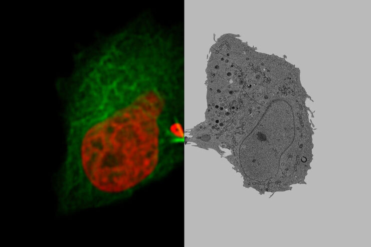
Putting Dynamic Live Cell Data into the Ultrastructural Context
With workflow Coral Life, searching for a needle in the haystack is a thing of the past. Take advantage of correlative light and electron microscopy to identify directly the right cell at the right…
