is mobile? false
Science Lab
Science Lab
Willkommen auf dem Wissensportal von Leica Microsystems. Hier finden Sie wissenschaftliches Forschungs- und Lehrmaterial rund um das Thema Mikroskopie. Das Portal unterstützt Anfänger, erfahrene Praktiker und Wissenschaftler gleichermaßen bei ihrer täglichen Arbeit und ihren Experimenten. Erkunden Sie interaktive Tutorials und Anwendungshinweise, entdecken Sie die Grundlagen der Mikroskopie ebenso wie High-End-Technologien. Werden Sie Teil der Science Lab Community und teilen Sie Ihr Fachwissen.
Wissensportal Show subnavigation

Science Lab
Wissens- und Lernportal für Mikroskopie. Lernen. Teilen. Mitmachen.


Wählen Sie Ihr Anwendungsgebiet Show subnavigation


Filter articles
Tags
Berichtstyp
Produkte
Loading...

Effiziente Aufnahme von langen Zeitserien
Bei Experimenten mit Aufnahmen langer Zeitserien von Sphäroiden können sich bestimmte Herausforderungen ergeben. Da die Experimente mehrere Tage dauern können, muss die Probe möglichst lange am Leben…
Loading...

Live-Cell Imaging Techniques
The understanding of complex and/or fast cellular dynamics is an important step for exploring biological processes. Therefore, today’s life science research is increasingly focused on dynamic…
Loading...

Imaging of Anti-Cancer Drug Uptake in Spheroids using DLS
Spheroid 3D cell culture models mimic the physiology and functions of living tissues making them a useful tool to study tumor morphology and screen anti-cancer drugs. The drug AZD2014 is a recognized…
Loading...
 in 3D")
How Artificial Intelligence Enhances Confocal Imaging
In this article, we show how artificial intelligence (AI) can enhance your imaging experiments. Namely, how Dynamic Signal Enhancement powered by Aivia improves image quality while capturing the…
Loading...

Benefits of Combining STED and Lifetime
In this interview, Professor Alberto Diaspro talks about the advantages of the White Light Laser and the TauSTED capabilities of STELLARIS 8 STED. He speaks about his experience with the confocal…
Loading...

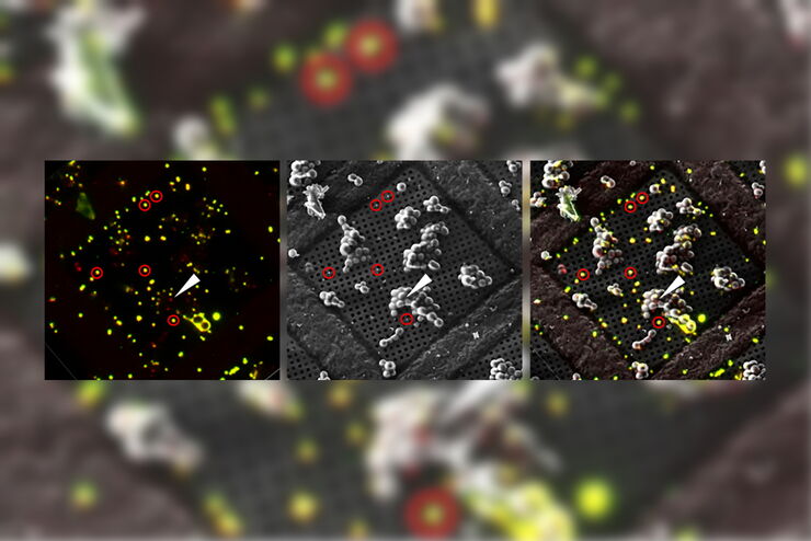
Spectroscopic Evaluation of Red Blood Cells
Hemoglobinopathies are a major healthcare problem. This study presents a possible diagnostic tool for thalassemia which is based on confocal spectroscopy. This approach exploits spectral detection and…
Loading...
The Cryo-CLEM Journey
This article describes the Cryo-CLEM technology and the benefits it can provide for scientists. Additionally, some scientific publications are highlighted.
Recent developments in cryo electron…
Loading...
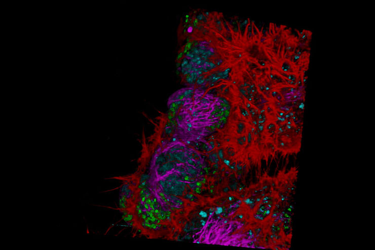
Fluorescence Lifetime-based Imaging Gallery
Confocal microscopy relies on the effective excitation of fluorescence probes and the efficient collection of photons emitted from the fluorescence process. One aspect of fluorescence is the emission…
Loading...
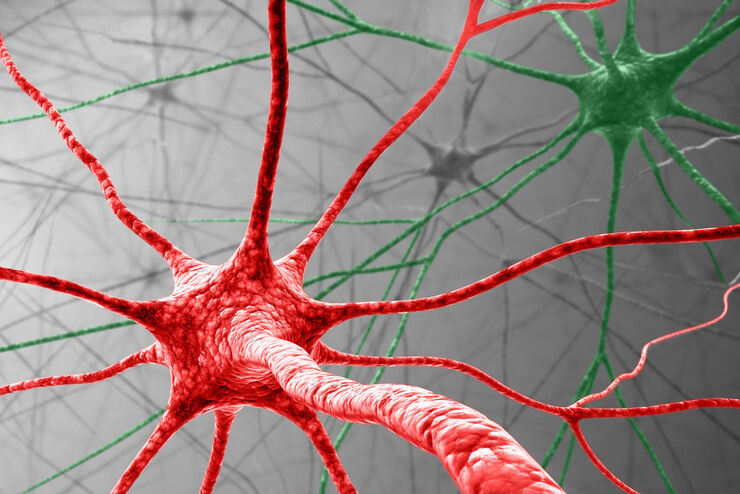
Visualizing Protein Degradation and Aggregation in the Living Cell
Our guest speaker, Prof Dr Eric Reits, presents his work on neurodegenerative disorders. Reits’ group are experts on the subject of Huntington’s disease and work towards identifying leads for…
