is mobile? false
Science Lab
Science Lab
Willkommen auf dem Wissensportal von Leica Microsystems. Hier finden Sie wissenschaftliches Forschungs- und Lehrmaterial rund um das Thema Mikroskopie. Das Portal unterstützt Anfänger, erfahrene Praktiker und Wissenschaftler gleichermaßen bei ihrer täglichen Arbeit und ihren Experimenten. Erkunden Sie interaktive Tutorials und Anwendungshinweise, entdecken Sie die Grundlagen der Mikroskopie ebenso wie High-End-Technologien. Werden Sie Teil der Science Lab Community und teilen Sie Ihr Fachwissen.
Wissensportal Show subnavigation

Science Lab
Wissens- und Lernportal für Mikroskopie. Lernen. Teilen. Mitmachen.


Wählen Sie Ihr Anwendungsgebiet Show subnavigation


Filter articles
Tags
Berichtstyp
Produkte
Loading...

Top Issues Related to Standards for Rating Non-Metallic Inclusions in Steel
Supplying components and products made of steel to users worldwide can require that a single batch be compliant with multiple steel quality standards. This user demand creates significant challenges…
Loading...

Analyzing Non-metallic Inclusions in Steel
Oftentimes we find ourselves caught up in tedious analyses by reticle and comparison chart, time-consuming double-evaluation according to several standards or subjective inspection results with a bias…
Loading...
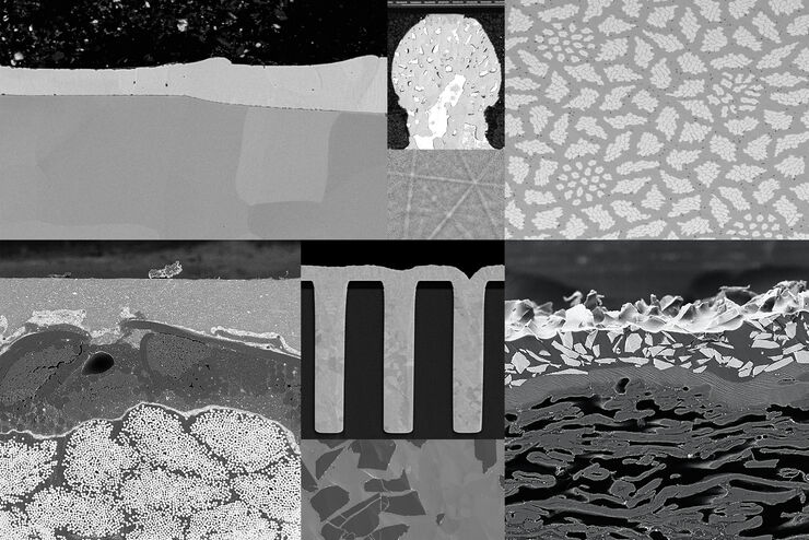
Introduction to Ion Beam Etching with the EM TIC 3X
In this article you can learn how to optimize the preparation quality of your samples by using the ion beam etching method with the EM TIC 3X ion beam milling machine. A short introduction of the…
Loading...

Rate the Quality of Your Steel: Free Webinar and Report
This webinar and report describe optimal microscopy solutions for rating steel quality in terms of non-metallic inclusions and reviews the various international and regional standards concerning…
Loading...
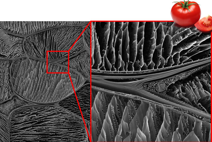
Studying the Microstructure of Natural Polymers in Fine Detail
The potential of cryogenic broad ion beam milling used in combination with scanning electron microscopy (cryo-BIB-SEM) for imaging and analyzing the microstructure of cryogenically stabilized soft…
Loading...
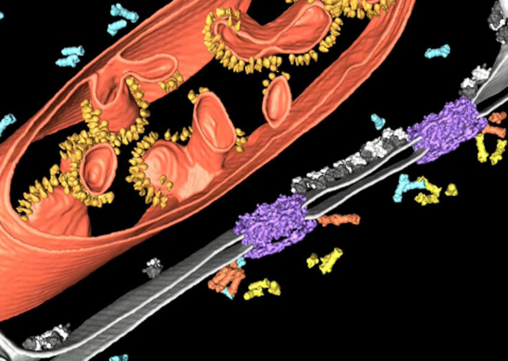
Improve Cryo Electron Tomography Workflow
Leica Microsystems and Thermo Fisher Scientific have collaborated to create a fully integrated cryo-tomography workflow that responds to these research needs: Reveal cellular mechanisms at…
Loading...
![3D glomeruli in a portion of an ECi-cleared kidney scanned by light sheet microscopy. Courtesy of Prof. Norbert Gretz, Medical Faculty Mannheim, University of Heidelberg [1].](/fileadmin/_processed_/d/d/csm_DLS-Sample-Preparation-Intr_915e0fd7c2.jpg "3D glomeruli in a portion of an ECi-cleared kidney scanned by light sheet microscopy. Courtesy of Prof. Norbert Gretz, Medical Faculty Mannheim, University of Heidelberg [1].")
Using Mounting Frames for Light Sheet Sample Preparation
Sample handling is an important topic in the context of Light Sheet Microscopy. The TCS SP8 DLS integrates Light Sheet technology into an inverted confocal platform and can hence make use of general…
Loading...

Using a Rotation Device for Light Sheet Sample Mounting
The TCS SP8 DLS from Leica Microsystems is an innovative concept to integrate the Light Sheet Microscopy technology into the confocal microscope. Due to its unique optical architecture samples can be…
Loading...

High Resolution Array Tomography with Automated Serial Sectioning
The optimization of high resolution, 3-dimensional (3D), sub-cellular structure analysis with array tomography using an automated serial sectioning solution, achieving a high section density on the…
